4GUU

Crystal structure of LSD2-NPAC with tranylcypromine


Experimental Data Snapshot

  • Method: X-RAY DIFFRACTION
  • Resolution: 2.30 Å
  • R-Value Free: 0.231 
  • R-Value Work: 0.199 
  • R-Value Observed: 0.200 

wwPDB Validation   3D Report Full Report


Ligand Structure Quality Assessment 


This is version 1.2 of the entry. See complete history


Literature

LSD2/KDM1B and its cofactor NPAC/GLYR1 endow a structural and molecular model for regulation of H3K4 demethylation

Fang, R.Chen, F.Dong, Z.Hu, D.Barbera, A.J.Clark, E.A.Fang, J.Yang, Y.Mei, P.Rutenberg, M.Li, Z.Zhang, Y.Xu, Y.Yang, H.Wang, P.Simon, M.D.Zhou, Q.Li, J.Marynick, M.P.Li, X.Lu, H.Kaiser, U.B.Kingston, R.E.Xu, Y.Shi, Y.G.

(2013) Mol Cell 49: 558-570

  • DOI: https://doi.org/10.1016/j.molcel.2012.11.019
  • Primary Citation of Related Structures:  
    4GU1, 4GUR, 4GUS, 4GUT, 4GUU

  • PubMed Abstract: 

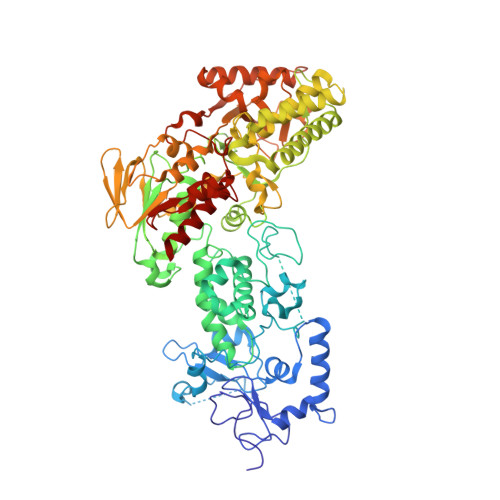
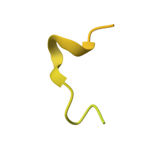
    Dynamic regulation of histone methylation represents a fundamental epigenetic mechanism underlying eukaryotic gene regulation, yet little is known about how the catalytic activities of histone demethylases are regulated. Here, we identify and characterize NPAC/GLYR1 as an LSD2/KDM1b-specific cofactor that stimulates H3K4me1 and H3K4me2 demethylation. We determine the crystal structures of LSD2 alone and LSD2 in complex with the NPAC linker region in the absence or presence of histone H3 peptide, at resolutions of 2.9, 2.0, and 2.25 Å, respectively. These crystal structures and further biochemical characterization define a dodecapeptide of NPAC (residues 214-225) as the minimal functional unit for its cofactor activity and provide structural determinants and a molecular mechanism underlying the intrinsic cofactor activity of NPAC in stimulating LSD2-catalyzed H3K4 demethylation. Thus, these findings establish a model for how a cofactor directly regulates histone demethylation and will have a significant impact on our understanding of catalytic-activity-based epigenetic regulation.


  • Organizational Affiliation

    Division of Endocrinology, Diabetes, and Hypertension, Department of Medicine and Department of Biological Chemistry & Molecular Pharmacology, Brigham and Women's Hospital and Harvard Medical School, 221 Longwood Avenue, Boston, MA 02115, USA.


Macromolecules
Find similar proteins by:  (by identity cutoff)  |  3D Structure
Entity ID: 1
MoleculeChains Sequence LengthOrganismDetailsImage
Lysine-specific histone demethylase 1B776Homo sapiensMutation(s): 0 
Gene Names: LSD2
EC: 1
UniProt & NIH Common Fund Data Resources
Find proteins for Q8NB78 (Homo sapiens)
Explore Q8NB78 
Go to UniProtKB:  Q8NB78
PHAROS:  Q8NB78
GTEx:  ENSG00000165097 
Entity Groups  
Sequence Clusters30% Identity50% Identity70% Identity90% Identity95% Identity100% Identity
UniProt GroupQ8NB78
Sequence Annotations
Expand
  • Reference Sequence
Find similar proteins by:  (by identity cutoff)  |  3D Structure
Entity ID: 2
MoleculeChains Sequence LengthOrganismDetailsImage
Putative oxidoreductase GLYR1124Homo sapiensMutation(s): 0 
Gene Names: GLYR1
EC: 1
UniProt & NIH Common Fund Data Resources
Find proteins for Q49A26 (Homo sapiens)
Explore Q49A26 
Go to UniProtKB:  Q49A26
PHAROS:  Q49A26
GTEx:  ENSG00000140632 
Entity Groups  
Sequence Clusters30% Identity50% Identity70% Identity90% Identity95% Identity100% Identity
UniProt GroupQ49A26
Sequence Annotations
Expand
  • Reference Sequence
Small Molecules
Ligands 2 Unique
IDChains Name / Formula / InChI Key2D Diagram3D Interactions
FA9
Query on FA9

Download Ideal Coordinates CCD File 
C [auth A][(2R,3S,4R,5R)-5-(6-amino-9H-purin-9-yl)-3,4-dihydroxytetrahydrofuran-2-yl]methyl (2R,3S,4S)-5-[(3R,3aS,7aR)-10,11-dimethyl-1,4,6-trioxo-3-phenyl-2,3,5,6,7,7a-hexahydro-1H-benzo[g]pyrrolo[2,1-e]pteridin-8(4H)-yl]-2,3,4-trihydroxypentyl dihydrogen diphosphate
C36 H43 N9 O16 P2
QWOOVYWBGZGGNV-ITRBMUPBSA-N
ZN
Query on ZN

Download Ideal Coordinates CCD File 
D [auth A],
E [auth A],
F [auth A]
ZINC ION
Zn
PTFCDOFLOPIGGS-UHFFFAOYSA-N
Experimental Data & Validation

Experimental Data

  • Method: X-RAY DIFFRACTION
  • Resolution: 2.30 Å
  • R-Value Free: 0.231 
  • R-Value Work: 0.199 
  • R-Value Observed: 0.200 
  • Space Group: P 1 21 1
Unit Cell:
Length ( Å )Angle ( ˚ )
a = 62.82α = 90
b = 90.157β = 104.2
c = 87.824γ = 90
Software Package:
Software NamePurpose
HKL-2000data collection
PHASERphasing
PHENIXrefinement
HKL-2000data reduction
HKL-2000data scaling

Structure Validation

View Full Validation Report


Ligand Structure Quality Assessment 


Entry History 

Deposition Data

Revision History  (Full details and data files)

  • Version 1.0: 2013-01-16
    Type: Initial release
  • Version 1.1: 2013-07-10
    Changes: Database references
  • Version 1.2: 2023-11-08
    Changes: Data collection, Database references, Derived calculations, Refinement description