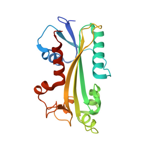
The human ITPA polymorphic variant P32T is destabilized by the unpacking of the hydrophobic core.
Simone, P.D., Struble, L.R., Kellezi, A., Brown, C.A., Grabow, C.E., Khutsishvili, I., Marky, L.A., Pavlov, Y.I., Borgstahl, G.E.(2013) J Struct Biol 182: 197-208
- PubMed: 23528839
- DOI: https://doi.org/10.1016/j.jsb.2013.03.007
- Primary Citation of Related Structures:
4F95 - PubMed Abstract:
Inosine triphosphate pyrophosphatase (ITPA), a key enzyme involved in maintaining the purity of cellular nucleoside triphosphate pools, specifically recognizes inosine triphosphate and xanthosine triphosphate (including the deoxyribose forms) and detoxifies them by catalyzing the hydrolysis of a phosphoanhydride bond, releasing pyrophosphate. This prevents their inappropriate use as substrates in enzymatic reactions utilizing (d)ATP or (d)GTP. A human genetic polymorphism leads to the substitution of Thr for Pro32 (P32T) and causes ITPA deficiency in erythrocytes, with heterozygotes having on average 22.5% residual activity, and homozygotes having undetectable activity. This polymorphism has been implicated in modulating patients' response to mercaptopurines and ribavirin. Human fibroblasts containing this variant have elevated genomic instability upon treatment with base analogs. We find that the wild-type and P32T forms are dimeric in solution and in the crystal structure. This abolishes the previous speculation that the P32T change disrupts dimerization as a mechanism of inactivation. The only difference in structure from the wild-type protein is that the area surrounding Thr32 is disrupted. Phe31 is flipped from the hydrophobic core out into the solvent, leaving a hole in the hydrophobic core of the protein which likely accounts for the reduced thermal stability of P32T ITPA and ultimately leads to its susceptibility to degradation in human cells. Circular dichroism and thermal denaturation studies confirm these structural results. We propose that the dimer of P32T variant subunit with wild-type subunit is degraded in cells similarly to the P32T homodimer explaining the level of loss of ITPA activity in heterozygotes.
Organizational Affiliation:
The Eppley Institute for Research in Cancer and Allied Diseases, University of Nebraska Medical Center, 987696 Nebraska Medical Center, Omaha, NE 68198-7696, USA.