4BUM

Crystal structure of the Voltage Dependant Anion Channel 2 from zebrafish.


Experimental Data Snapshot

  • Method: X-RAY DIFFRACTION
  • Resolution: 2.80 Å
  • R-Value Free: 0.282 
  • R-Value Work: 0.245 
  • R-Value Observed: 0.249 

wwPDB Validation   3D Report Full Report


Ligand Structure Quality Assessment 


This is version 1.3 of the entry. See complete history


Literature

High-Resolution Structure and Double Electron-Electron Resonance of the Zebrafish Voltage Dependent Anion Channel 2 Reveal an Oligomeric Population.

Schredelseker, J.Paz, A.Lopez, C.J.Altenbach, C.Leung, C.S.Drexler, M.K.Chen, J.Hubbell, W.L.Abramson, J.

(2014) J Biol Chem 289: 12566

  • DOI: https://doi.org/10.1074/jbc.M113.497438
  • Primary Citation of Related Structures:  
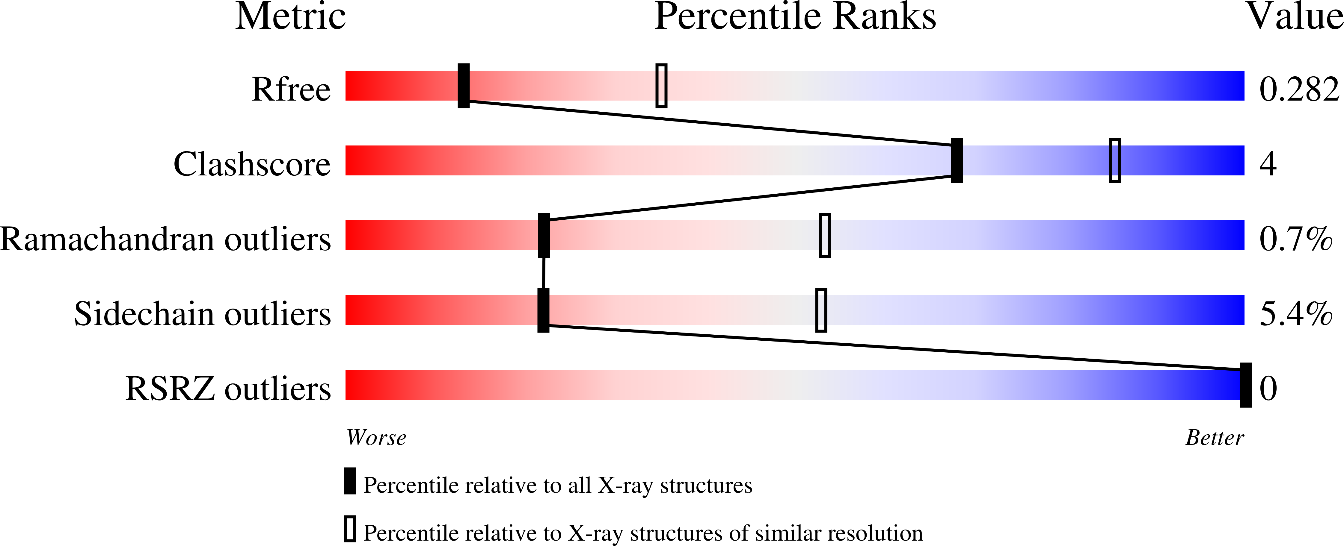
    4BUM

  • PubMed Abstract: 

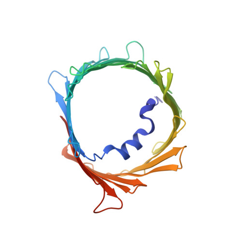
    In recent years, there has been a vast increase in structural and functional understanding of VDAC1, but VDAC2 and -3 have been understudied despite having many unique phenotypes. One reason for the paucity of structural and biochemical characterization of the VDAC2 and -3 isoforms stems from the inability of obtaining purified, functional protein. Here we demonstrate the expression, isolation, and basic characterization of zebrafish VDAC2 (zfVDAC2). Further, we resolved the structure of zfVDAC2 at 2.8 Å resolution, revealing a crystallographic dimer. The dimer orientation was confirmed in solution by double electron-electron resonance spectroscopy and by cross-linking experiments disclosing a dimer population of ∼20% in lauryldimethine amine oxide detergent micelles, whereas in lipidic bicelles a higher population of dimeric and higher order oligomers species were observed. The present study allows for a more accurate structural comparison between VDAC2 and its better-studied counterpart VDAC1.


  • Organizational Affiliation

    From the Department of Molecular, Cell and Developmental Biology.


Macromolecules
Find similar proteins by:  (by identity cutoff)  |  3D Structure
Entity ID: 1
MoleculeChains Sequence LengthOrganismDetailsImage
VOLTAGE-DEPENDENT ANION CHANNEL 2A [auth X]289Danio rerioMutation(s): 0 
Membrane Entity: Yes 
UniProt
Find proteins for Q8AWD0 (Danio rerio)
Explore Q8AWD0 
Go to UniProtKB:  Q8AWD0
Entity Groups  
Sequence Clusters30% Identity50% Identity70% Identity90% Identity95% Identity100% Identity
UniProt GroupQ8AWD0
Sequence Annotations
Expand
  • Reference Sequence
Small Molecules
Ligands 1 Unique
IDChains Name / Formula / InChI Key2D Diagram3D Interactions
LDA
Query on LDA

Download Ideal Coordinates CCD File 
B [auth X]LAURYL DIMETHYLAMINE-N-OXIDE
C14 H31 N O
SYELZBGXAIXKHU-UHFFFAOYSA-N
Experimental Data & Validation

Experimental Data

  • Method: X-RAY DIFFRACTION
  • Resolution: 2.80 Å
  • R-Value Free: 0.282 
  • R-Value Work: 0.245 
  • R-Value Observed: 0.249 
  • Space Group: P 32 2 1
Unit Cell:
Length ( Å )Angle ( ˚ )
a = 72.47α = 90
b = 72.47β = 90
c = 177.95γ = 120
Software Package:
Software NamePurpose
PHENIXrefinement
PHENIXphasing

Structure Validation

View Full Validation Report


Ligand Structure Quality Assessment 


Entry History 

Deposition Data

Revision History  (Full details and data files)

  • Version 1.0: 2014-03-26
    Type: Initial release
  • Version 1.1: 2014-04-09
    Changes: Structure summary
  • Version 1.2: 2014-05-14
    Changes: Database references
  • Version 1.3: 2023-12-20
    Changes: Data collection, Database references, Derived calculations, Other, Refinement description