4ZNE

IgG1 Fc-FcgammaRI ecd complex


Experimental Data Snapshot

  • Method: X-RAY DIFFRACTION
  • Resolution: 2.42 Å
  • R-Value Free: 0.254 
  • R-Value Work: 0.201 
  • R-Value Observed: 0.203 

wwPDB Validation   3D Report Full Report


Ligand Structure Quality Assessment 


This is version 2.0 of the entry. See complete history


Literature

Structural insights into the interaction of human IgG1 with Fc gamma RI: no direct role of glycans in binding.

Oganesyan, V.Mazor, Y.Yang, C.Cook, K.E.Woods, R.M.Ferguson, A.Bowen, M.A.Martin, T.Zhu, J.Wu, H.Dall'Acqua, W.F.

(2015) Acta Crystallogr D Biol Crystallogr 71: 2354-2361

  • DOI: https://doi.org/10.1107/S1399004715018015
  • Primary Citation of Related Structures:  
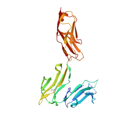
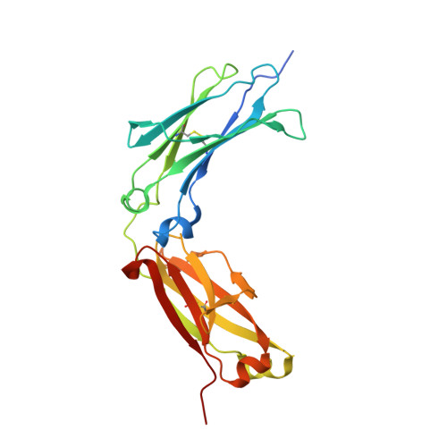
    4ZNE

  • PubMed Abstract: 

    The three-dimensional structure of a human IgG1 Fc fragment bound to wild-type human FcγRI is reported. The structure of the corresponding complex was solved at a resolution of 2.4 Å using molecular replacement; this is the highest resolution achieved for an unmutated FcγRI molecule. This study highlights the critical structural and functional role played by the second extracellular subdomain of FcγRI. It also explains the long-known major energetic contribution of the Fc `LLGG' motif at positions 234-237, and particularly of Leu235, via a `lock-and-key' mechanism. Finally, a previously held belief is corrected and a differing view is offered on the recently proposed direct role of Fc carbohydrates in the corresponding interaction. Structural evidence is provided that such glycan-related effects are strictly indirect.


  • Organizational Affiliation

    Department of Antibody Discovery and Protein Engineering, MedImmune LLC, 1 MedImmune Way, Gaithersburg, MD 20878, USA.


Macromolecules
Find similar proteins by:  (by identity cutoff)  |  3D Structure
Entity ID: 1
MoleculeChains Sequence LengthOrganismDetailsImage
High affinity immunoglobulin gamma Fc receptor I267Homo sapiensMutation(s): 0 
Gene Names: FCGR1AFCG1FCGR1IGFR1
UniProt & NIH Common Fund Data Resources
Find proteins for P12314 (Homo sapiens)
Explore P12314 
Go to UniProtKB:  P12314
PHAROS:  P12314
GTEx:  ENSG00000150337 
Entity Groups  
Sequence Clusters30% Identity50% Identity70% Identity90% Identity95% Identity100% Identity
UniProt GroupP12314
Sequence Annotations
Expand
  • Reference Sequence
Find similar proteins by:  (by identity cutoff)  |  3D Structure
Entity ID: 2
MoleculeChains Sequence LengthOrganismDetailsImage
Ig gamma-1 chain C regionB [auth E],
C [auth J]
227Homo sapiensMutation(s): 0 
Gene Names: IGHG1
UniProt & NIH Common Fund Data Resources
Find proteins for P01857 (Homo sapiens)
Explore P01857 
Go to UniProtKB:  P01857
PHAROS:  P01857
GTEx:  ENSG00000211896 
Entity Groups  
Sequence Clusters30% Identity50% Identity70% Identity90% Identity95% Identity100% Identity
UniProt GroupP01857
Sequence Annotations
Expand
  • Reference Sequence
Oligosaccharides

Help

Entity ID: 3
MoleculeChains Length2D Diagram Glycosylation3D Interactions
alpha-D-mannopyranose-(1-3)-2-acetamido-2-deoxy-beta-D-glucopyranose-(1-4)-2-acetamido-2-deoxy-beta-D-glucopyranoseD [auth B]3N-Glycosylation
Glycosylation Resources
GlyTouCan:  G66019PC
GlyCosmos:  G66019PC
GlyGen:  G66019PC
Entity ID: 4
MoleculeChains Length2D Diagram Glycosylation3D Interactions
alpha-D-mannopyranose-(1-4)-2-acetamido-2-deoxy-beta-D-glucopyranoseE [auth C]2N-Glycosylation
Glycosylation Resources
GlyTouCan:  G35893LO
GlyCosmos:  G35893LO
GlyGen:  G35893LO
Entity ID: 5
MoleculeChains Length2D Diagram Glycosylation3D Interactions
2-acetamido-2-deoxy-beta-D-glucopyranose-(1-4)-2-acetamido-2-deoxy-beta-D-glucopyranoseF [auth D]2N-Glycosylation
Glycosylation Resources
GlyTouCan:  G42666HT
GlyCosmos:  G42666HT
GlyGen:  G42666HT
Entity ID: 6
MoleculeChains Length2D Diagram Glycosylation3D Interactions
2-acetamido-2-deoxy-beta-D-glucopyranose-(1-2)-alpha-D-mannopyranose-(1-3)-[2-acetamido-2-deoxy-beta-D-glucopyranose-(1-2)-alpha-D-mannopyranose-(1-6)]alpha-D-mannopyranose-(1-4)-2-acetamido-2-deoxy-beta-D-glucopyranose-(1-4)-[beta-L-fucopyranose-(1-6)]2-acetamido-2-deoxy-beta-D-glucopyranoseG [auth F],
H [auth G]
8N-Glycosylation
Glycosylation Resources
GlyTouCan:  G88372NG
GlyCosmos:  G88372NG
GlyGen:  G88372NG
Experimental Data & Validation

Experimental Data

  • Method: X-RAY DIFFRACTION
  • Resolution: 2.42 Å
  • R-Value Free: 0.254 
  • R-Value Work: 0.201 
  • R-Value Observed: 0.203 
  • Space Group: C 1 2 1
Unit Cell:
Length ( Å )Angle ( ˚ )
a = 134.735α = 90
b = 126.75β = 118.44
c = 71.844γ = 90
Software Package:
Software NamePurpose
REFMACrefinement
XDSdata reduction
XDSdata scaling
MOLREPphasing

Structure Validation

View Full Validation Report


Ligand Structure Quality Assessment 


Entry History 

Deposition Data

Revision History  (Full details and data files)

  • Version 1.0: 2015-11-11
    Type: Initial release
  • Version 1.1: 2015-11-18
    Changes: Database references
  • Version 2.0: 2020-07-29
    Type: Remediation
    Reason: Carbohydrate remediation
    Changes: Advisory, Atomic model, Data collection, Derived calculations, Structure summary