4ZFQ

Structure of M. tuberculosis (3,3) L,D-Transpeptidase, LdtMt5. (Meropenen-adduct form)


Experimental Data Snapshot

  • Method: X-RAY DIFFRACTION
  • Resolution: 2.80 Å
  • R-Value Free: 0.277 
  • R-Value Work: 0.230 
  • R-Value Observed: 0.232 

wwPDB Validation   3D Report Full Report


Ligand Structure Quality Assessment 


This is version 1.4 of the entry. See complete history


Literature

Loss of a Functionally and Structurally Distinct ld-Transpeptidase, LdtMt5, Compromises Cell Wall Integrity in Mycobacterium tuberculosis.

Brammer Basta, L.A.Ghosh, A.Pan, Y.Jakoncic, J.Lloyd, E.P.Townsend, C.A.Lamichhane, G.Bianchet, M.A.

(2015) J Biol Chem 290: 25670-25685

  • DOI: https://doi.org/10.1074/jbc.M115.660753
  • Primary Citation of Related Structures:  
    4Z7A, 4ZFQ

  • PubMed Abstract: 

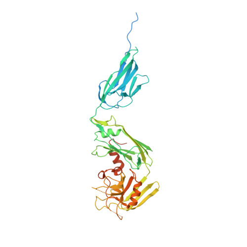
    The final step of peptidoglycan (PG) biosynthesis in bacteria involves cross-linking of peptide side chains. This step in Mycobacterium tuberculosis is catalyzed by ld- and dd-transpeptidases that generate 3→3 and 4→3 transpeptide linkages, respectively. M. tuberculosis PG is predominantly 3→3 cross-linked, and LdtMt2 is the dominant ld-transpeptidase. There are four additional sequence paralogs of LdtMt2 encoded by the genome of this pathogen, and the reason for this apparent redundancy is unknown. Here, we studied one of the paralogs, LdtMt5, and found it to be structurally and functionally distinct. The structures of apo-LdtMt5 and its meropenem adduct presented here demonstrate that, despite overall architectural similarity to LdtMt2, the LdtMt5 active site has marked differences. The presence of a structurally divergent catalytic site and a proline-rich C-terminal subdomain suggest that this protein may have a distinct role in PG metabolism, perhaps involving other cell wall-anchored proteins. Furthermore, M. tuberculosis lacking a functional copy of LdtMt5 displayed aberrant growth and was more susceptible to killing by crystal violet, osmotic shock, and select carbapenem antibiotics. Therefore, we conclude that LdtMt5 is not a functionally redundant ld-transpeptidase, but rather it serves a unique and important role in maintaining the integrity of the M. tuberculosis cell wall.


  • Organizational Affiliation

    From the Taskforce to study Resistance Emergence and Antimicrobial development Technology (TREAT) and Division of Infectious Diseases, The Johns Hopkins University School of Medicine, Baltimore, Maryland 21231.


Macromolecules
Find similar proteins by:  (by identity cutoff)  |  3D Structure
Entity ID: 1
MoleculeChains Sequence LengthOrganismDetailsImage
L,D-transpeptidase 5451Mycobacterium tuberculosis CDC1551Mutation(s): 0 
Gene Names: MT0501
EC: 2.3.2
UniProt
Find proteins for P9WKV2 (Mycobacterium tuberculosis (strain CDC 1551 / Oshkosh))
Explore P9WKV2 
Go to UniProtKB:  P9WKV2
Entity Groups  
Sequence Clusters30% Identity50% Identity70% Identity90% Identity95% Identity100% Identity
UniProt GroupP9WKV2
Sequence Annotations
Expand
  • Reference Sequence
Small Molecules
Ligands 2 Unique
IDChains Name / Formula / InChI Key2D Diagram3D Interactions
DWZ
Query on DWZ

Download Ideal Coordinates CCD File 
D [auth A](2S,3R,4S)-4-{[(3S,5S)-5-(dimethylcarbamoyl)pyrrolidin-3-yl]sulfanyl}-2-[(2S,3R)-3-hydroxy-1-oxobutan-2-yl]-3-methyl-3,4-dihydro-2H-pyrrole-5-carboxylic acid
C17 H27 N3 O5 S
UUIYVKJXUXGPKB-VGWSNGFZSA-N
PEG
Query on PEG

Download Ideal Coordinates CCD File 
B [auth A],
C [auth A]
DI(HYDROXYETHYL)ETHER
C4 H10 O3
MTHSVFCYNBDYFN-UHFFFAOYSA-N
Experimental Data & Validation

Experimental Data

  • Method: X-RAY DIFFRACTION
  • Resolution: 2.80 Å
  • R-Value Free: 0.277 
  • R-Value Work: 0.230 
  • R-Value Observed: 0.232 
  • Space Group: P 62 2 2
Unit Cell:
Length ( Å )Angle ( ˚ )
a = 99.393α = 90
b = 99.393β = 90
c = 193.384γ = 120
Software Package:
Software NamePurpose
PHENIXrefinement
HKL-2000data reduction
SCALEPACKdata scaling
MOLREPphasing

Structure Validation

View Full Validation Report


Ligand Structure Quality Assessment 


Entry History 

Deposition Data

Revision History  (Full details and data files)

  • Version 1.0: 2015-09-02
    Type: Initial release
  • Version 1.1: 2015-09-09
    Changes: Database references
  • Version 1.2: 2015-10-28
    Changes: Database references
  • Version 1.3: 2022-02-02
    Changes: Database references, Derived calculations, Structure summary
  • Version 1.4: 2023-09-27
    Changes: Data collection, Refinement description