4X4M

Structure of FcgammaRI in complex with Fc reveals the importance of glycan recognition for high affinity IgG binding


Experimental Data Snapshot

  • Method: X-RAY DIFFRACTION
  • Resolution: 3.48 Å
  • R-Value Free: 0.296 
  • R-Value Work: 0.245 
  • R-Value Observed: 0.249 

wwPDB Validation   3D Report Full Report


Ligand Structure Quality Assessment 


This is version 2.0 of the entry. See complete history


Literature

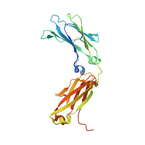
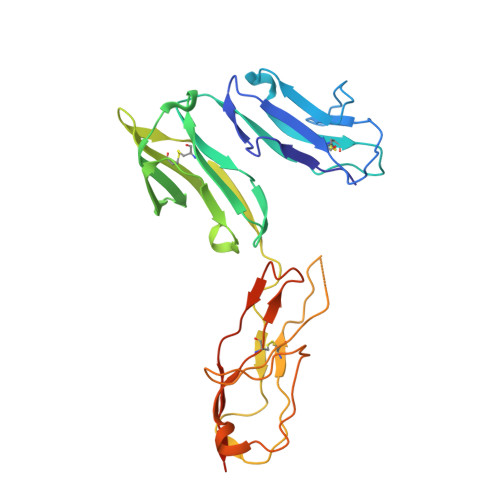
Structure of Fc gamma RI in complex with Fc reveals the importance of glycan recognition for high-affinity IgG binding.

Lu, J.Chu, J.Zou, Z.Hamacher, N.B.Rixon, M.W.Sun, P.D.

(2015) Proc Natl Acad Sci U S A 112: 833-838

  • DOI: https://doi.org/10.1073/pnas.1418812112
  • Primary Citation of Related Structures:  
    4X4M

  • PubMed Abstract: 

    Fc gamma receptor I (FcγRI) contributes to protective immunity against bacterial infections, but exacerbates certain autoimmune diseases. The sole high-affinity IgG receptor, FcγRI plays a significant role in immunotherapy. To elucidate the molecular mechanism of its high-affinity IgG binding, we determined the crystal structure of the extracellular domains of human FcγRI in complex with the Fc domain of human IgG1. FcγRI binds to the Fc in a similar mode as the low-affinity FcγRII and FcγRIII receptors. In addition to many conserved contacts, FcγRI forms additional hydrogen bonds and salt bridges with the lower hinge region of Fc. Unique to the high-affinity receptor-Fc complex, however, is the conformation of the receptor D2 domain FG loop, which enables a charged KHR motif to interact with proximal carbohydrate units of the Fc glycans. Both the length and the charge of the FcγRI FG loop are well conserved among mammalian species. Ala and Glu mutations of the FG loop KHR residues showed significant contributions of His-174 and Arg-175 to antibody binding, and the loss of the FG loop-glycan interaction resulted in an ∼ 20- to 30-fold decrease in FcγRI affinity to all three subclasses of IgGs. Furthermore, deglycosylation of IgG1 resulted in a 40-fold loss in FcγRI binding, demonstrating involvement of the receptor FG loop in glycan recognition. These results highlight a unique glycan recognition in FcγRI function and open potential therapeutic avenues based on antibody glycan engineering or small molecular glycan mimics to target FcγRI for certain autoimmune diseases.


  • Organizational Affiliation

    Structural Immunology Section, Laboratory of Immunogenetics, National Institute of Allergy and Infectious Diseases, National Institutes of Health, Rockville, MD 20852 and.


Macromolecules
Find similar proteins by:  (by identity cutoff)  |  3D Structure
Entity ID: 1
MoleculeChains Sequence LengthOrganismDetailsImage
Ig gamma-1 chain C region
A, B, C, D
219Homo sapiensMutation(s): 0 
Gene Names: IGHG1
UniProt & NIH Common Fund Data Resources
Find proteins for P01857 (Homo sapiens)
Explore P01857 
Go to UniProtKB:  P01857
PHAROS:  P01857
GTEx:  ENSG00000211896 
Entity Groups  
Sequence Clusters30% Identity50% Identity70% Identity90% Identity95% Identity100% Identity
UniProt GroupP01857
Sequence Annotations
Expand
  • Reference Sequence
Find similar proteins by:  (by identity cutoff)  |  3D Structure
Entity ID: 2
MoleculeChains Sequence LengthOrganismDetailsImage
High affinity immunoglobulin gamma Fc receptor I
E, F
275Homo sapiensMutation(s): 18 
Gene Names: FCGR1AFCG1FCGR1IGFR1
UniProt & NIH Common Fund Data Resources
Find proteins for P12314 (Homo sapiens)
Explore P12314 
Go to UniProtKB:  P12314
PHAROS:  P12314
GTEx:  ENSG00000150337 
Entity Groups  
Sequence Clusters30% Identity50% Identity70% Identity90% Identity95% Identity100% Identity
UniProt GroupP12314
Sequence Annotations
Expand
  • Reference Sequence
Oligosaccharides

Help

Entity ID: 3
MoleculeChains Length2D Diagram Glycosylation3D Interactions
2-acetamido-2-deoxy-beta-D-glucopyranose-(1-2)-alpha-D-mannopyranose-(1-3)-[2-acetamido-2-deoxy-beta-D-glucopyranose-(1-2)-alpha-D-mannopyranose-(1-6)]beta-D-mannopyranose-(1-4)-2-acetamido-2-deoxy-beta-D-glucopyranose-(1-4)-[beta-L-fucopyranose-(1-6)]2-acetamido-2-deoxy-beta-D-glucopyranose
G, H
8N-Glycosylation
Glycosylation Resources
GlyTouCan:  G59451NL
GlyCosmos:  G59451NL
GlyGen:  G59451NL
Entity ID: 4
MoleculeChains Length2D Diagram Glycosylation3D Interactions
beta-D-galactopyranose-(1-4)-2-acetamido-2-deoxy-beta-D-glucopyranose-(1-2)-alpha-D-mannopyranose-(1-6)-[2-acetamido-2-deoxy-beta-D-glucopyranose-(1-2)-alpha-D-mannopyranose-(1-3)]beta-D-mannopyranose-(1-4)-2-acetamido-2-deoxy-beta-D-glucopyranose-(1-4)-[beta-L-fucopyranose-(1-6)]2-acetamido-2-deoxy-beta-D-glucopyranose
I
9N-Glycosylation
Glycosylation Resources
GlyTouCan:  G33244HE
GlyCosmos:  G33244HE
GlyGen:  G33244HE
Entity ID: 5
MoleculeChains Length2D Diagram Glycosylation3D Interactions
2-acetamido-2-deoxy-beta-D-glucopyranose-(1-2)-alpha-D-mannopyranose-(1-3)-[2-acetamido-2-deoxy-beta-D-glucopyranose-(1-2)-alpha-D-mannopyranose-(1-6)]beta-D-mannopyranose-(1-4)-2-acetamido-2-deoxy-beta-D-glucopyranose
J
6N/A
Glycosylation Resources
GlyTouCan:  G55061AW
GlyCosmos:  G55061AW
GlyGen:  G55061AW
Entity ID: 6
MoleculeChains Length2D Diagram Glycosylation3D Interactions
N-acetyl-alpha-neuraminic acid-(2-3)-beta-D-galactopyranose
K
2N/A
Glycosylation Resources
GlyTouCan:  G30207PZ
GlyCosmos:  G30207PZ
GlyGen:  G30207PZ
Small Molecules
Ligands 2 Unique
IDChains Name / Formula / InChI Key2D Diagram3D Interactions
NAG
Query on NAG

Download Ideal Coordinates CCD File 
L [auth D]2-acetamido-2-deoxy-beta-D-glucopyranose
C8 H15 N O6
OVRNDRQMDRJTHS-FMDGEEDCSA-N
FUL
Query on FUL

Download Ideal Coordinates CCD File 
M [auth D]beta-L-fucopyranose
C6 H12 O5
SHZGCJCMOBCMKK-KGJVWPDLSA-N
Experimental Data & Validation

Experimental Data

  • Method: X-RAY DIFFRACTION
  • Resolution: 3.48 Å
  • R-Value Free: 0.296 
  • R-Value Work: 0.245 
  • R-Value Observed: 0.249 
  • Space Group: P 1
Unit Cell:
Length ( Å )Angle ( ˚ )
a = 59.921α = 89.86
b = 67.991β = 112.25
c = 125.028γ = 89.96
Software Package:
Software NamePurpose
PHENIXrefinement
HKL-2000data reduction
SCALAdata scaling
PHASERphasing

Structure Validation

View Full Validation Report


Ligand Structure Quality Assessment 


Entry History 

Deposition Data

Revision History  (Full details and data files)

  • Version 1.0: 2015-04-08
    Type: Initial release
  • Version 2.0: 2020-07-29
    Type: Remediation
    Reason: Carbohydrate remediation
    Changes: Advisory, Atomic model, Data collection, Database references, Derived calculations, Source and taxonomy, Structure summary