4WD3

Crystal structure of an L-amino acid ligase RizA


Experimental Data Snapshot

  • Method: X-RAY DIFFRACTION
  • Resolution: 2.80 Å
  • R-Value Free: 0.255 
  • R-Value Work: 0.201 
  • R-Value Observed: 0.204 

wwPDB Validation   3D Report Full Report


This is version 1.1 of the entry. See complete history


Literature

Structure of RizA, an L-amino-acid ligase from Bacillus subtilis.

Kagawa, W.Arai, T.Ishikura, S.Kino, K.Kurumizaka, H.

(2015) Acta Crystallogr F Struct Biol Commun 71: 1125-1130

  • DOI: https://doi.org/10.1107/S2053230X15012698
  • Primary Citation of Related Structures:  
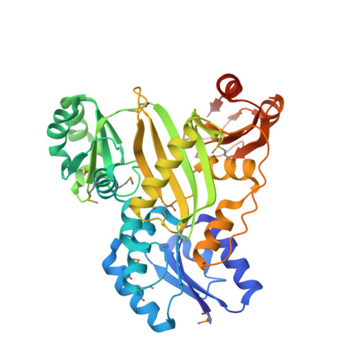
    4WD3

  • PubMed Abstract: 

    RizA is an L-amino-acid ligase from Bacillus subtilis that participates in the biosynthesis of rhizocticin, an oligopeptide antibiotic. The substrate-free form of RizA has been crystallized and the structure was solved at 2.8 Å resolution. The amino-acid-binding site appears to be capable of accommodating multiple amino acids, consistent with previous biochemical studies.


  • Organizational Affiliation

    Department of Interdisciplinary Science and Engineering, Program in Chemistry and Life Science, School of Science and Engineering, Meisei University, 2-1-1 Hodokubo, Hino-shi, Tokyo 191-8506, Japan.


Macromolecules
Find similar proteins by:  (by identity cutoff)  |  3D Structure
Entity ID: 1
MoleculeChains Sequence LengthOrganismDetailsImage
L-amino acid ligase
A, B
413Bacillus subtilis subsp. subtilisMutation(s): 2 
Gene Names: rizA
UniProt
Find proteins for B5UAT8 (Bacillus subtilis)
Explore B5UAT8 
Go to UniProtKB:  B5UAT8
Entity Groups  
Sequence Clusters30% Identity50% Identity70% Identity90% Identity95% Identity100% Identity
UniProt GroupB5UAT8
Sequence Annotations
Expand
  • Reference Sequence
Small Molecules
Modified Residues  1 Unique
IDChains TypeFormula2D DiagramParent
MSE
Query on MSE
A, B
L-PEPTIDE LINKINGC5 H11 N O2 SeMET
Experimental Data & Validation

Experimental Data

  • Method: X-RAY DIFFRACTION
  • Resolution: 2.80 Å
  • R-Value Free: 0.255 
  • R-Value Work: 0.201 
  • R-Value Observed: 0.204 
  • Space Group: P 1 21 1
Unit Cell:
Length ( Å )Angle ( ˚ )
a = 58.38α = 90
b = 87.29β = 97.52
c = 85.26γ = 90
Software Package:
Software NamePurpose
PHENIXrefinement

Structure Validation

View Full Validation Report


Entry History & Funding Information

Deposition Data


Funding OrganizationLocationGrant Number
Japan--

Revision History  (Full details and data files)

  • Version 1.0: 2015-09-09
    Type: Initial release
  • Version 1.1: 2020-01-01
    Changes: Data collection, Database references, Derived calculations