4UP2

Crystal structure of Escherichia coli tryptophanase purified from alkaline stressed bacterial culture.


Experimental Data Snapshot

  • Method: X-RAY DIFFRACTION
  • Resolution: 2.78 Å
  • R-Value Free: 0.246 
  • R-Value Work: 0.215 
  • R-Value Observed: 0.217 

wwPDB Validation   3D Report Full Report


This is version 2.1 of the entry. See complete history


Literature

Structure of Escherichia Coli Tryptophanase Purified from an Alkaline-Stressed Bacterial Culture.

Rety, S.Deschamps, P.Leulliot, N.

(2015) Acta Crystallogr F Struct Biol Commun 71: 1378

  • DOI: https://doi.org/10.1107/S2053230X15017549
  • Primary Citation of Related Structures:  
    4UP2

  • PubMed Abstract: 

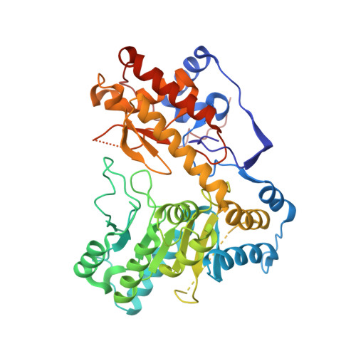
    Tryptophanase is a bacterial enzyme involved in the degradation of tryptophan to indole, pyruvate and ammonia, which are compounds that are essential for bacterial survival. Tryptophanase is often overexpressed in stressed cultures. Large amounts of endogenous tryptophanase were purified from Escherichia coli BL21 strain overexpressing another recombinant protein. Tryptophanase was crystallized in space group P6522 in the apo form without pyridoxal 5'-phosphate bound in the active site.


  • Organizational Affiliation

    Laboratoire de Cristallographie et RMN Biologiques, UMR CNRS 8015, Université Paris Descartes, Sorbonne Paris Cité, Faculté de Pharmacie de Paris, Paris, France.


Macromolecules
Find similar proteins by:  (by identity cutoff)  |  3D Structure
Entity ID: 1
MoleculeChains Sequence LengthOrganismDetailsImage
TRYPTOPHANASE
A, B, C, D
471Escherichia coliMutation(s): 0 
EC: 4.1.99.1
UniProt
Find proteins for C3SLI7 (Escherichia coli)
Explore C3SLI7 
Go to UniProtKB:  C3SLI7
Entity Groups  
Sequence Clusters30% Identity50% Identity70% Identity90% Identity95% Identity100% Identity
UniProt GroupC3SLI7
Sequence Annotations
Expand
  • Reference Sequence
Small Molecules
Ligands 2 Unique
IDChains Name / Formula / InChI Key2D Diagram3D Interactions
SO4
Query on SO4

Download Ideal Coordinates CCD File 
AA [auth C]
BA [auth D]
CA [auth D]
DA [auth D]
E [auth A]
AA [auth C],
BA [auth D],
CA [auth D],
DA [auth D],
E [auth A],
EA [auth D],
F [auth A],
FA [auth D],
G [auth A],
GA [auth D],
H [auth A],
I [auth A],
J [auth A],
K [auth B],
L [auth B],
M [auth B],
N [auth B],
O [auth B],
P [auth B],
Q [auth B],
S [auth B],
T [auth C],
U [auth C],
V [auth C],
W [auth C],
X [auth C],
Y [auth C],
Z [auth C]
SULFATE ION
O4 S
QAOWNCQODCNURD-UHFFFAOYSA-L
BME
Query on BME

Download Ideal Coordinates CCD File 
R [auth B]BETA-MERCAPTOETHANOL
C2 H6 O S
DGVVWUTYPXICAM-UHFFFAOYSA-N
Experimental Data & Validation

Experimental Data

  • Method: X-RAY DIFFRACTION
  • Resolution: 2.78 Å
  • R-Value Free: 0.246 
  • R-Value Work: 0.215 
  • R-Value Observed: 0.217 
  • Space Group: P 65 2 2
Unit Cell:
Length ( Å )Angle ( ˚ )
a = 158.24α = 90
b = 158.24β = 90
c = 387.766γ = 120
Software Package:
Software NamePurpose
PHENIXrefinement
XDSdata reduction
XDSdata scaling
PHASERphasing

Structure Validation

View Full Validation Report


Entry History 

Deposition Data

Revision History  (Full details and data files)

  • Version 1.0: 2015-06-24
    Type: Initial release
  • Version 1.1: 2015-11-11
    Changes: Database references
  • Version 2.0: 2019-10-23
    Changes: Atomic model, Data collection, Other
  • Version 2.1: 2024-01-10
    Changes: Data collection, Database references, Derived calculations, Refinement description