


4UIP
The complex structure of extracellular domain of EGFR with Repebody (rAC1).
- PDB DOI: https://doi.org/10.2210/pdb4UIP/pdb
- Classification: TRANSFERASE
- Organism(s): Homo sapiens, Listeria monocytogenes, synthetic construct, Eptatretus burgeri
- Expression System: Spodoptera frugiperda, Escherichia coli
- Mutation(s): No
- Deposited: 2015-03-31 Released: 2015-11-25
Experimental Data Snapshot
- Method: X-RAY DIFFRACTION
- Resolution: 2.95 Å
- R-Value Free: 0.297
- R-Value Work: 0.237
- R-Value Observed: 0.240

This is version 2.1 of the entry. See complete history.

Macromolecules
Find similar proteins by:
(by identity cutoff) | 3D Structure
Entity ID: 1 | |||||
|---|---|---|---|---|---|
| Molecule | Chains | Sequence Length | Organism | Details | Image |
| EPIDERMAL GROWTH FACTOR RECEPTOR | 618 | Homo sapiens | Mutation(s): 0 | 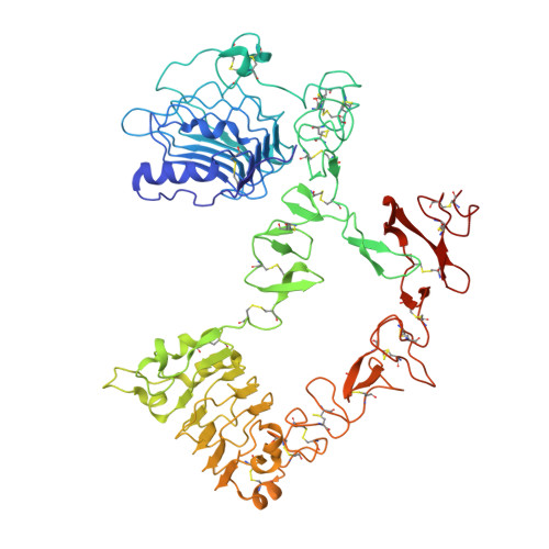 | |
UniProt & NIH Common Fund Data Resources | |||||
Find proteins for P00533 (Homo sapiens) Explore P00533 Go to UniProtKB: P00533 | |||||
PHAROS: P00533 GTEx: ENSG00000146648 | |||||
Entity Groups | |||||
| Sequence Clusters | 30% Identity50% Identity70% Identity90% Identity95% Identity100% Identity | ||||
| UniProt Group | P00533 | ||||
Sequence AnnotationsExpand | |||||
| |||||
Find similar proteins by:
(by identity cutoff) | 3D Structure
Entity ID: 2 | |||||
|---|---|---|---|---|---|
| Molecule | Chains | Sequence Length | Organism | Details | Image |
| REPEBODY (RAC1) | 251 | Listeria monocytogenes, synthetic construct, Eptatretus burgeri This entity is chimeric | Mutation(s): 0 |  | |
UniProt | |||||
Find proteins for Q4G1L3 (Eptatretus burgeri) Explore Q4G1L3 Go to UniProtKB: Q4G1L3 | |||||
Find proteins for E0ACT6 (Listeria monocytogenes) Explore E0ACT6 Go to UniProtKB: E0ACT6 | |||||
Entity Groups | |||||
| Sequence Clusters | 30% Identity50% Identity70% Identity90% Identity95% Identity100% Identity | ||||
| UniProt Groups | E0ACT6Q4G1L3 | ||||
Sequence AnnotationsExpand | |||||
| |||||
Oligosaccharides

Small Molecules
| Ligands 1 Unique | |||||
|---|---|---|---|---|---|
| ID | Chains | Name / Formula / InChI Key | 2D Diagram | 3D Interactions | |
| NAG Query on NAG | D [auth A], E [auth A] | 2-acetamido-2-deoxy-beta-D-glucopyranose C8 H15 N O6 OVRNDRQMDRJTHS-FMDGEEDCSA-N |  | ||
Experimental Data & Validation
Experimental Data
- Method: X-RAY DIFFRACTION
- Resolution: 2.95 Å
- R-Value Free: 0.297
- R-Value Work: 0.237
- R-Value Observed: 0.240
- Space Group: P 2 21 21
Unit Cell:
| Length ( Å ) | Angle ( ˚ ) |
|---|---|
| a = 66.79 | α = 90 |
| b = 88.34 | β = 90 |
| c = 189.21 | γ = 90 |
| Software Name | Purpose |
|---|---|
| REFMAC | refinement |
Entry History
Deposition Data
- Released Date: 2015-11-25 Deposition Author(s): Kang, Y.J., Cha, Y.J., Cho, H.S., Lee, J.J., Kim, H.S.
Revision History (Full details and data files)
- Version 1.0: 2015-11-25
Type: Initial release - Version 1.1: 2015-12-23
Changes: Structure summary - Version 1.2: 2017-03-15
Changes: Source and taxonomy - Version 2.0: 2020-07-29
Type: Remediation
Reason: Carbohydrate remediation
Changes: Advisory, Atomic model, Data collection, Derived calculations, Other, Structure summary - Version 2.1: 2024-01-10
Changes: Data collection, Database references, Refinement description, Structure summary
