4UF7

Ghanaian henipavirus (Gh-M74a) attachment glycoprotein in complex with human ephrinB2


Experimental Data Snapshot

  • Method: X-RAY DIFFRACTION
  • Resolution: 1.70 Å
  • R-Value Free: 0.199 
  • R-Value Work: 0.175 
  • R-Value Observed: 0.176 

wwPDB Validation   3D Report Full Report


Ligand Structure Quality Assessment 


This is version 1.5 of the entry. See complete history


Literature

Molecular Recognition of Human Ephrinb2 Cell Surface Receptor by an Emergent African Henipavirus.

Lee, B.Pernet, O.Ahmed, A.A.Zeltina, A.Beaty, S.M.Bowden, T.A.

(2015) Proc Natl Acad Sci U S A 112: E2156

  • DOI: https://doi.org/10.1073/pnas.1501690112
  • Primary Citation of Related Structures:  
    4UF7

  • PubMed Abstract: 

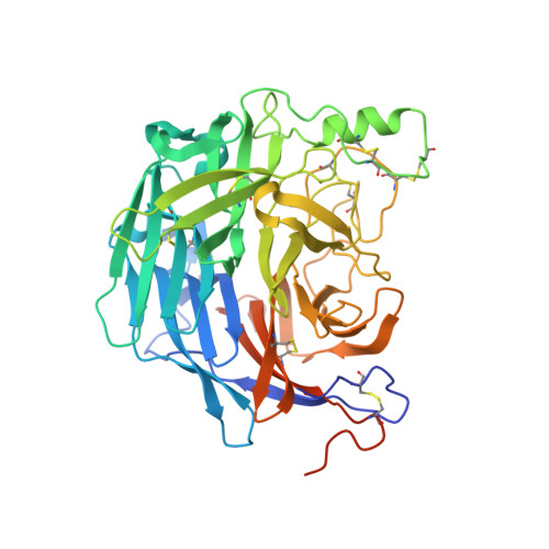
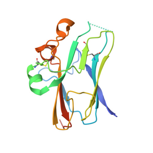
    The discovery of African henipaviruses (HNVs) related to pathogenic Hendra virus (HeV) and Nipah virus (NiV) from Southeast Asia and Australia presents an open-ended health risk. Cell receptor use by emerging African HNVs at the stage of host-cell entry is a key parameter when considering the potential for spillover and infection of human populations. The attachment glycoprotein from a Ghanaian bat isolate (GhV-G) exhibits <30% sequence identity with Asiatic NiV-G/HeV-G. Here, through functional and structural analysis of GhV-G, we show how this African HNV targets the same human cell-surface receptor (ephrinB2) as the Asiatic HNVs. We first characterized this virus-receptor interaction crystallographically. Compared with extant HNV-G-ephrinB2 structures, there was significant structural variation in the six-bladed β-propeller scaffold of the GhV-G receptor-binding domain, but not the Greek key fold of the bound ephrinB2. Analysis revealed a surprisingly conserved mode of ephrinB2 interaction that reflects an ongoing evolutionary constraint among geographically distal and phylogenetically divergent HNVs to maintain the functionality of ephrinB2 recognition during virus-host entry. Interestingly, unlike NiV-G/HeV-G, we could not detect binding of GhV-G to ephrinB3. Comparative structure-function analysis further revealed several distinguishing features of HNV-G function: a secondary ephrinB2 interaction site that contributes to more efficient ephrinB2-mediated entry in NiV-G relative to GhV-G and cognate residues at the very C terminus of GhV-G (absent in Asiatic HNV-Gs) that are vital for efficient receptor-induced fusion, but not receptor binding per se. These data provide molecular-level details for evaluating the likelihood of African HNVs to spill over into human populations.


  • Organizational Affiliation

    Department of Microbiology, Icahn School of Medicine at Mount Sinai, New York, NY 10029; thomas.bowden@strubi.ox.ac.uk benhur.lee@mssm.edu.


Macromolecules
Find similar proteins by:  (by identity cutoff)  |  3D Structure
Entity ID: 1
MoleculeChains Sequence LengthOrganismDetailsImage
GLYCOPROTEIN
A, B
454Ghana virusMutation(s): 0 
UniProt
Find proteins for I0E093 (Ghana virus)
Explore I0E093 
Go to UniProtKB:  I0E093
Entity Groups  
Sequence Clusters30% Identity50% Identity70% Identity90% Identity95% Identity100% Identity
UniProt GroupI0E093
Sequence Annotations
Expand
  • Reference Sequence
Find similar proteins by:  (by identity cutoff)  |  3D Structure
Entity ID: 2
MoleculeChains Sequence LengthOrganismDetailsImage
EPHRIN-B2C,
D [auth E]
153Homo sapiensMutation(s): 0 
UniProt & NIH Common Fund Data Resources
Find proteins for P52799 (Homo sapiens)
Explore P52799 
Go to UniProtKB:  P52799
PHAROS:  P52799
GTEx:  ENSG00000125266 
Entity Groups  
Sequence Clusters30% Identity50% Identity70% Identity90% Identity95% Identity100% Identity
UniProt GroupP52799
Sequence Annotations
Expand
  • Reference Sequence
Small Molecules
Ligands 4 Unique
IDChains Name / Formula / InChI Key2D Diagram3D Interactions
NAG
Query on NAG

Download Ideal Coordinates CCD File 
CA [auth B]
DA [auth B]
EA [auth B]
FA [auth B]
HA [auth C]
CA [auth B],
DA [auth B],
EA [auth B],
FA [auth B],
HA [auth C],
JA [auth E],
KA [auth E],
P [auth A],
Q [auth A],
R [auth A],
S [auth A]
2-acetamido-2-deoxy-beta-D-glucopyranose
C8 H15 N O6
OVRNDRQMDRJTHS-FMDGEEDCSA-N
SO4
Query on SO4

Download Ideal Coordinates CCD File 
E [auth A]
F [auth A]
G [auth A]
GA [auth C]
H [auth A]
E [auth A],
F [auth A],
G [auth A],
GA [auth C],
H [auth A],
I [auth A],
J [auth A],
T [auth B],
U [auth B],
V [auth B],
W [auth B],
X [auth B],
Y [auth B]
SULFATE ION
O4 S
QAOWNCQODCNURD-UHFFFAOYSA-L
ACT
Query on ACT

Download Ideal Coordinates CCD File 
AA [auth B]
IA [auth E]
K [auth A]
L [auth A]
M [auth A]
AA [auth B],
IA [auth E],
K [auth A],
L [auth A],
M [auth A],
N [auth A],
Z [auth B]
ACETATE ION
C2 H3 O2
QTBSBXVTEAMEQO-UHFFFAOYSA-M
CL
Query on CL

Download Ideal Coordinates CCD File 
BA [auth B],
O [auth A]
CHLORIDE ION
Cl
VEXZGXHMUGYJMC-UHFFFAOYSA-M
Experimental Data & Validation

Experimental Data

  • Method: X-RAY DIFFRACTION
  • Resolution: 1.70 Å
  • R-Value Free: 0.199 
  • R-Value Work: 0.175 
  • R-Value Observed: 0.176 
  • Space Group: I 2 2 2
Unit Cell:
Length ( Å )Angle ( ˚ )
a = 127.22α = 90
b = 152.53β = 90
c = 163.3γ = 90
Software Package:
Software NamePurpose
REFMACrefinement
xia2data reduction
xia2data scaling
PHASERphasing

Structure Validation

View Full Validation Report


Ligand Structure Quality Assessment 


Entry History 

Deposition Data

Revision History  (Full details and data files)

  • Version 1.0: 2015-04-01
    Type: Initial release
  • Version 1.1: 2015-04-15
    Changes: Database references
  • Version 1.2: 2015-05-13
    Changes: Database references
  • Version 1.3: 2018-02-28
    Changes: Source and taxonomy
  • Version 1.4: 2020-07-29
    Type: Remediation
    Reason: Carbohydrate remediation
    Changes: Data collection, Derived calculations, Other, Structure summary
  • Version 1.5: 2023-12-20
    Changes: Data collection, Database references, Refinement description, Structure summary