4QLC

Crystal structure of chromatosome at 3.5 angstrom resolution


Experimental Data Snapshot

  • Method: X-RAY DIFFRACTION
  • Resolution: 3.50 Å
  • R-Value Free: 0.243 
  • R-Value Work: 0.218 
  • R-Value Observed: 0.220 

wwPDB Validation   3D Report Full Report


This is version 1.4 of the entry. See complete history


Literature

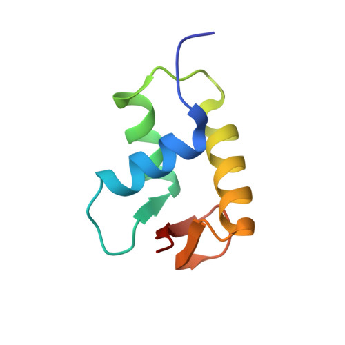
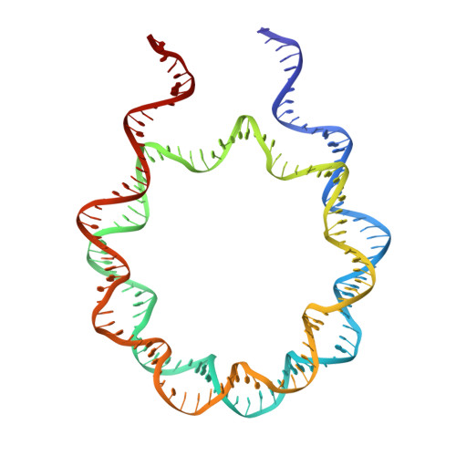
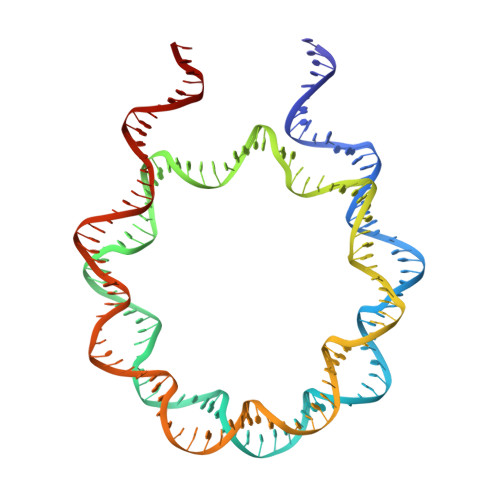
Structural Mechanisms of Nucleosome Recognition by Linker Histones.

Zhou, B.R.Jiang, J.Feng, H.Ghirlando, R.Xiao, T.S.Bai, Y.

(2015) Mol Cell 33 Suppl 1: 2-3

  • DOI: https://doi.org/10.1016/j.molcel.2015.06.025
  • Primary Citation of Related Structures:  
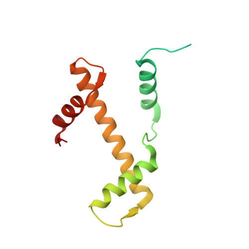
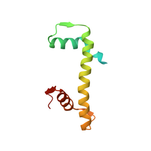
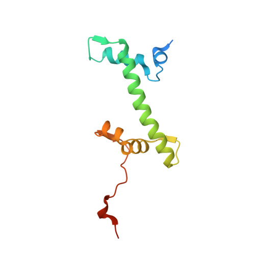
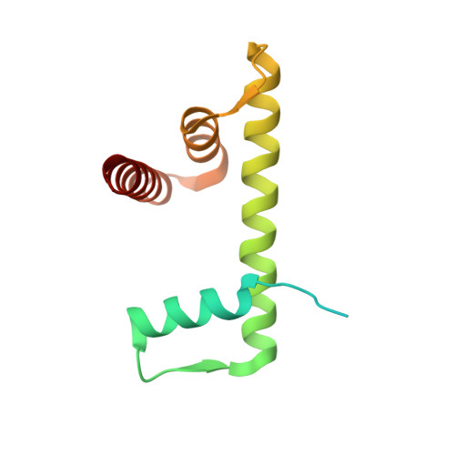
    4QLC

  • PubMed Abstract: 

    Linker histones bind to the nucleosome and regulate the structure of chromatin and gene expression. Despite more than three decades of effort, the structural basis of nucleosome recognition by linker histones remains elusive. Here, we report the crystal structure of the globular domain of chicken linker histone H5 in complex with the nucleosome at 3.5 Å resolution, which is validated using nuclear magnetic resonance spectroscopy. The globular domain sits on the dyad of the nucleosome and interacts with both DNA linkers. Our structure integrates results from mutation analyses and previous cross-linking and fluorescence recovery after photobleach experiments, and it helps resolve the long debate on structural mechanisms of nucleosome recognition by linker histones. The on-dyad binding mode of the H5 globular domain is different from the recently reported off-dyad binding mode of Drosophila linker histone H1. We demonstrate that linker histones with different binding modes could fold chromatin to form distinct higher-order structures.


  • Organizational Affiliation

    Laboratory of Biochemistry and Molecular Biology, National Cancer Institute, National Institutes of Health, Bethesda, MD 20892, USA.


Macromolecules

Find similar proteins by:  (by identity cutoff)  |  3D Structure
Entity ID: 3
MoleculeChains Sequence LengthOrganismDetailsImage
Histone H3C [auth A],
G [auth E]
135Drosophila melanogasterMutation(s): 0 
Gene Names: 
UniProt
Find proteins for P02299 (Drosophila melanogaster)
Explore P02299 
Go to UniProtKB:  P02299
Entity Groups  
Sequence Clusters30% Identity50% Identity70% Identity90% Identity95% Identity100% Identity
UniProt GroupP02299
Sequence Annotations
Expand
  • Reference Sequence
Find similar proteins by:  (by identity cutoff)  |  3D Structure
Entity ID: 4
MoleculeChains Sequence LengthOrganismDetailsImage
Histone H4D [auth B],
H [auth F]
102Drosophila melanogasterMutation(s): 0 
Gene Names: 
UniProt
Find proteins for P84040 (Drosophila melanogaster)
Explore P84040 
Go to UniProtKB:  P84040
Entity Groups  
Sequence Clusters30% Identity50% Identity70% Identity90% Identity95% Identity100% Identity
UniProt GroupP84040
Sequence Annotations
Expand
  • Reference Sequence
Find similar proteins by:  (by identity cutoff)  |  3D Structure
Entity ID: 5
MoleculeChains Sequence LengthOrganismDetailsImage
Histone H2AE [auth C],
I [auth G]
123Drosophila melanogasterMutation(s): 0 
Gene Names: 
UniProt
Find proteins for P84051 (Drosophila melanogaster)
Explore P84051 
Go to UniProtKB:  P84051
Entity Groups  
Sequence Clusters30% Identity50% Identity70% Identity90% Identity95% Identity100% Identity
UniProt GroupP84051
Sequence Annotations
Expand
  • Reference Sequence
Find similar proteins by:  (by identity cutoff)  |  3D Structure
Entity ID: 6
MoleculeChains Sequence LengthOrganismDetailsImage
Histone H2BF [auth D],
J [auth H]
122Drosophila melanogasterMutation(s): 0 
Gene Names: 
UniProt
Find proteins for P02283 (Drosophila melanogaster)
Explore P02283 
Go to UniProtKB:  P02283
Entity Groups  
Sequence Clusters30% Identity50% Identity70% Identity90% Identity95% Identity100% Identity
UniProt GroupP02283
Sequence Annotations
Expand
  • Reference Sequence
Find similar proteins by:  (by identity cutoff)  |  3D Structure
Entity ID: 7
MoleculeChains Sequence LengthOrganismDetailsImage
H5K [auth U]77Gallus gallusMutation(s): 0 
Gene Names: H5_CHICK
UniProt
Find proteins for P02259 (Gallus gallus)
Explore P02259 
Go to UniProtKB:  P02259
Entity Groups  
Sequence Clusters30% Identity50% Identity70% Identity90% Identity95% Identity100% Identity
UniProt GroupP02259
Sequence Annotations
Expand
  • Reference Sequence
Find similar nucleic acids by:  (by identity cutoff)  |  3D Structure
Entity ID: 1
MoleculeChains LengthOrganismImage
DNA (167-mer)A [auth I]167Homo sapiens
Sequence Annotations
Expand
  • Reference Sequence
Find similar nucleic acids by:  (by identity cutoff)  |  3D Structure
Entity ID: 2
MoleculeChains LengthOrganismImage
DNA (167-mer)B [auth J]167Homo sapiens
Sequence Annotations
Expand
  • Reference Sequence
Experimental Data & Validation

Experimental Data

  • Method: X-RAY DIFFRACTION
  • Resolution: 3.50 Å
  • R-Value Free: 0.243 
  • R-Value Work: 0.218 
  • R-Value Observed: 0.220 
  • Space Group: P 65
Unit Cell:
Length ( Å )Angle ( ˚ )
a = 262.87α = 90
b = 262.87β = 90
c = 91.79γ = 120
Software Package:
Software NamePurpose
Blu-Icedata collection
PHASERphasing
PHENIXrefinement
XDSdata reduction
XDSdata scaling

Structure Validation

View Full Validation Report


Entry History 

Deposition Data

Revision History  (Full details and data files)

  • Version 1.0: 2015-07-22
    Type: Initial release
  • Version 1.1: 2015-07-29
    Changes: Database references, Derived calculations
  • Version 1.2: 2015-08-12
    Changes: Database references
  • Version 1.3: 2017-11-22
    Changes: Advisory, Refinement description
  • Version 1.4: 2023-09-20
    Changes: Advisory, Data collection, Database references, Derived calculations, Refinement description