4Q0A

Vitamin D Receptor complex with lithocholic acid


Experimental Data Snapshot

  • Method: X-RAY DIFFRACTION
  • Resolution: 1.90 Å
  • R-Value Free: 0.244 
  • R-Value Work: 0.213 
  • R-Value Observed: 0.213 

wwPDB Validation   3D Report Full Report


Ligand Structure Quality Assessment 


This is version 1.2 of the entry. See complete history


Literature

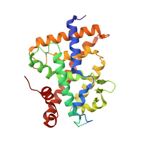
Structural insights into the molecular mechanism of vitamin d receptor activation by lithocholic Acid involving a new mode of ligand recognition.

Belorusova, A.Y.Eberhardt, J.Potier, N.Stote, R.H.Dejaegere, A.Rochel, N.

(2014) J Med Chem 57: 4710-4719

  • DOI: https://doi.org/10.1021/jm5002524
  • Primary Citation of Related Structures:  
    4Q0A

  • PubMed Abstract: 

    The vitamin D receptor (VDR), an endocrine nuclear receptor for 1α,25-dihydroxyvitamin D3, acts also as a bile acid sensor by binding lithocholic acid (LCA). The crystal structure of the zebrafish VDR ligand binding domain in complex with LCA and the SRC-2 coactivator peptide reveals the binding of two LCA molecules by VDR. One LCA binds to the canonical ligand-binding pocket, and the second one, which is not fully buried, is anchored to a site located on the VDR surface. Despite the low affinity of the alternative site, the binding of the second molecule promotes stabilization of the active receptor conformation. Biological activity assays, structural analysis, and molecular dynamics simulations indicate that the recognition of two ligand molecules is crucial for VDR agonism by LCA. The unique binding mode of LCA provides clues for the development of new chemical compounds that target alternative binding sites for therapeutic applications.


  • Organizational Affiliation

    Department of Integrative Structural Biology, Institut de Génétique et de Biologie Moléculaire et Cellulaire (IGBMC), Institut National de Santé et de Recherche Médicale (INSERM) U964, Centre National de Recherche Scientifique (CNRS) UMR 7104, Université de Strasbourg, 67404 Illkirch, France.


Macromolecules
Find similar proteins by:  (by identity cutoff)  |  3D Structure
Entity ID: 1
MoleculeChains Sequence LengthOrganismDetailsImage
Vitamin D3 receptor AA [auth C]302Danio rerioMutation(s): 0 
Gene Names: vdranr1i1avdr
UniProt
Find proteins for Q9PTN2 (Danio rerio)
Explore Q9PTN2 
Go to UniProtKB:  Q9PTN2
Entity Groups  
Sequence Clusters30% Identity50% Identity70% Identity90% Identity95% Identity100% Identity
UniProt GroupQ9PTN2
Sequence Annotations
Expand
  • Reference Sequence

Find similar proteins by:  Sequence   |   3D Structure  

Entity ID: 2
MoleculeChains Sequence LengthOrganismDetailsImage
Nuclear receptor coactivator 2B [auth D]10Homo sapiensMutation(s): 0 
UniProt & NIH Common Fund Data Resources
Find proteins for Q15596 (Homo sapiens)
Explore Q15596 
Go to UniProtKB:  Q15596
PHAROS:  Q15596
GTEx:  ENSG00000140396 
Entity Groups  
Sequence Clusters30% Identity50% Identity70% Identity90% Identity95% Identity100% Identity
UniProt GroupQ15596
Sequence Annotations
Expand
  • Reference Sequence
Small Molecules
Ligands 1 Unique
IDChains Name / Formula / InChI Key2D Diagram3D Interactions
4OA
Query on 4OA

Download Ideal Coordinates CCD File 
C,
D [auth C]
(3beta,5beta,14beta,17alpha)-3-hydroxycholan-24-oic acid
C24 H40 O3
SMEROWZSTRWXGI-HVATVPOCSA-N
Binding Affinity Annotations 
IDSourceBinding Affinity
4OA Binding MOAD:  4Q0A Kd: 330 (nM) from 1 assay(s)
Experimental Data & Validation

Experimental Data

  • Method: X-RAY DIFFRACTION
  • Resolution: 1.90 Å
  • R-Value Free: 0.244 
  • R-Value Work: 0.213 
  • R-Value Observed: 0.213 
  • Space Group: P 65 2 2
Unit Cell:
Length ( Å )Angle ( ˚ )
a = 66.213α = 90
b = 66.213β = 90
c = 262.571γ = 120
Software Package:
Software NamePurpose
HKL-2000data collection
CNSrefinement
HKL-2000data reduction
HKL-2000data scaling
CNSphasing

Structure Validation

View Full Validation Report


Ligand Structure Quality Assessment 


Entry History 

Deposition Data

Revision History  (Full details and data files)

  • Version 1.0: 2014-07-02
    Type: Initial release
  • Version 1.1: 2021-03-10
    Changes: Database references, Derived calculations
  • Version 1.2: 2024-02-28
    Changes: Data collection, Database references