4PCW

Crystal Structure of the N-terminal Domain of Human Profilaggrin at 2.2 A Resolution


Experimental Data Snapshot

  • Method: X-RAY DIFFRACTION
  • Resolution: 2.20 Å
  • R-Value Free: 0.242 
  • R-Value Work: 0.196 
  • R-Value Observed: 0.198 

wwPDB Validation   3D Report Full Report


This is version 1.3 of the entry. See complete history


Literature

Crystal Structure of Human Profilaggrin S100 Domain and Identification of Target Proteins Annexin II, Stratifin, and HSP27.

Bunick, C.G.Presland, R.B.Lawrence, O.T.Pearton, D.J.Milstone, L.M.Steitz, T.A.

(2015) J Invest Dermatol 135: 1801-1809

  • DOI: https://doi.org/10.1038/jid.2015.102
  • Primary Citation of Related Structures:  
    4PCW

  • PubMed Abstract: 

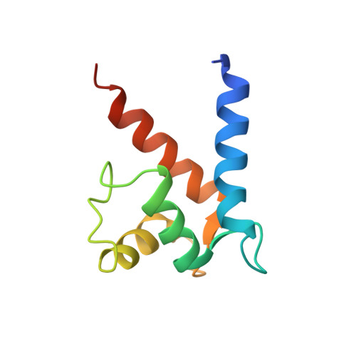
    The fused-type S100 protein profilaggrin and its proteolytic products including filaggrin are important in the formation of a normal epidermal barrier; however, the specific function of the S100 calcium-binding domain in profilaggrin biology is poorly understood. To explore its molecular function, we determined a 2.2 Å-resolution crystal structure of the N-terminal fused-type S100 domain of human profilaggrin with bound calcium ions. The profilaggrin S100 domain formed a stable dimer, which contained two hydrophobic pockets that provide a molecular interface for protein interactions. Biochemical and molecular approaches demonstrated that three proteins, annexin II/p36, stratifin/14-3-3 sigma, and heat shock protein 27, bind to the N-terminal domain of human profilaggrin; one protein (stratifin) co-localized with profilaggrin in the differentiating granular cell layer of human skin. Together, these findings suggest a model where the profilaggrin N-terminus uses calcium-dependent and calcium-independent protein-protein interactions to regulate its involvement in keratinocyte terminal differentiation and incorporation into the cornified cell envelope.


  • Organizational Affiliation

    Department of Dermatology, Yale University, New Haven, Connecticut, USA; Department of Molecular Biophysics and Biochemistry, Yale University, New Haven, Connecticut, USA; The first two authors contributed equally to this work.. Electronic address: christopher.bunick@yale.edu.


Macromolecules
Find similar proteins by:  (by identity cutoff)  |  3D Structure
Entity ID: 1
MoleculeChains Sequence LengthOrganismDetailsImage
Filaggrin
A, B, C, D
94Homo sapiensMutation(s): 0 
Gene Names: FLG
UniProt & NIH Common Fund Data Resources
Find proteins for P20930 (Homo sapiens)
Explore P20930 
Go to UniProtKB:  P20930
PHAROS:  P20930
GTEx:  ENSG00000143631 
Entity Groups  
Sequence Clusters30% Identity50% Identity70% Identity90% Identity95% Identity100% Identity
UniProt GroupP20930
Sequence Annotations
Expand
  • Reference Sequence
Small Molecules
Ligands 2 Unique
IDChains Name / Formula / InChI Key2D Diagram3D Interactions
2PE
Query on 2PE

Download Ideal Coordinates CCD File 
G [auth A],
L [auth C]
NONAETHYLENE GLYCOL
C18 H38 O10
YZUUTMGDONTGTN-UHFFFAOYSA-N
CA
Query on CA

Download Ideal Coordinates CCD File 
E [auth A]
F [auth A]
H [auth B]
I [auth B]
J [auth C]
E [auth A],
F [auth A],
H [auth B],
I [auth B],
J [auth C],
K [auth C],
M [auth D],
N [auth D]
CALCIUM ION
Ca
BHPQYMZQTOCNFJ-UHFFFAOYSA-N
Experimental Data & Validation

Experimental Data

  • Method: X-RAY DIFFRACTION
  • Resolution: 2.20 Å
  • R-Value Free: 0.242 
  • R-Value Work: 0.196 
  • R-Value Observed: 0.198 
  • Space Group: P 21 21 21
Unit Cell:
Length ( Å )Angle ( ˚ )
a = 40.492α = 90
b = 85.285β = 90
c = 96.901γ = 90
Software Package:
Software NamePurpose
PHENIXrefinement
HKL-2000data scaling
MOLREPphasing

Structure Validation

View Full Validation Report


Entry History & Funding Information

Deposition Data


Funding OrganizationLocationGrant Number
Dermatology FoundationUnited StatesCareer Development Award

Revision History  (Full details and data files)

  • Version 1.0: 2015-04-01
    Type: Initial release
  • Version 1.1: 2015-06-24
    Changes: Database references
  • Version 1.2: 2017-11-22
    Changes: Derived calculations, Other, Refinement description, Source and taxonomy
  • Version 1.3: 2023-09-27
    Changes: Data collection, Database references, Derived calculations, Refinement description