Structural basis of the heterodimerization of the MST and RASSF SARAH domains in the Hippo signalling pathway.
Hwang, E., Cheong, H.K., Mushtaq, A.U., Kim, H.Y., Yeo, K.J., Kim, E., Lee, W.C., Hwang, K.Y., Cheong, C., Jeon, Y.H.(2014) Acta Crystallogr D Biol Crystallogr 70: 1944-1953
- PubMed: 25004971
- DOI: https://doi.org/10.1107/S139900471400947X
- Primary Citation of Related Structures:
4OH8, 4OH9 - PubMed Abstract:
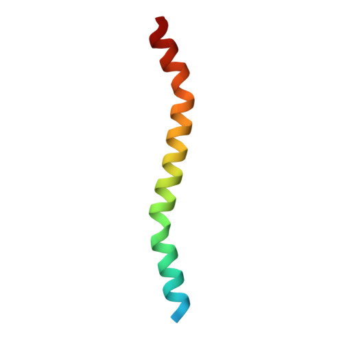
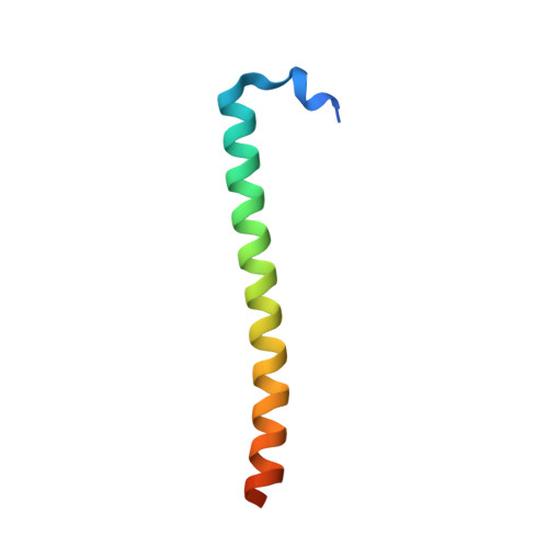
Despite recent progress in research on the Hippo signalling pathway, the structural information available in this area is extremely limited. Intriguingly, the homodimeric and heterodimeric interactions of mammalian sterile 20-like (MST) kinases through the so-called `SARAH' (SAV/RASSF/HPO) domains play a critical role in cellular homeostasis, dictating the fate of the cell regarding cell proliferation or apoptosis. To understand the mechanism of the heterodimerization of SARAH domains, the three-dimensional structures of an MST1-RASSF5 SARAH heterodimer and an MST2 SARAH homodimer were determined by X-ray crystallography and were analysed together with that previously determined for the MST1 SARAH homodimer. While the structure of the MST2 homodimer resembled that of the MST1 homodimer, the MST1-RASSF5 heterodimer showed distinct structural features. Firstly, the six N-terminal residues (Asp432-Lys437), which correspond to the short N-terminal 3₁₀-helix h1 kinked from the h2 helix in the MST1 homodimer, were disordered. Furthermore, the MST1 SARAH domain in the MST1-RASSF5 complex showed a longer helical structure (Ser438-Lys480) than that in the MST1 homodimer (Val441-Lys480). Moreover, extensive polar and nonpolar contacts in the MST1-RASSF5 SARAH domain were identified which strengthen the interactions in the heterodimer in comparison to the interactions in the homodimer. Denaturation experiments performed using urea also indicated that the MST-RASSF heterodimers are substantially more stable than the MST homodimers. These findings provide structural insights into the role of the MST1-RASSF5 SARAH domain in apoptosis signalling.
Organizational Affiliation:
Division of Magnetic Resonance Research, Korea Basic Science Institute, Ochang-eup Yeongudangiro 162, Cheongwon-gun, Chungbuk 363-883, Republic of Korea.