4NUF

Crystal Structure of SHP/EID1


Experimental Data Snapshot

  • Method: X-RAY DIFFRACTION
  • Resolution: 2.80 Å
  • R-Value Free: 0.245 
  • R-Value Work: 0.196 
  • R-Value Observed: 0.196 

wwPDB Validation   3D Report Full Report


This is version 2.1 of the entry. See complete history


Literature

Structural insights into gene repression by the orphan nuclear receptor SHP.

Zhi, X.Zhou, X.E.He, Y.Zechner, C.Suino-Powell, K.M.Kliewer, S.A.Melcher, K.Mangelsdorf, D.J.Xu, H.E.

(2014) Proc Natl Acad Sci U S A 111: 839-844

  • DOI: https://doi.org/10.1073/pnas.1322827111
  • Primary Citation of Related Structures:  
    4NUF

  • PubMed Abstract: 

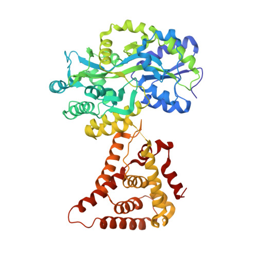
    Small heterodimer partner (SHP) is an orphan nuclear receptor that functions as a transcriptional repressor to regulate bile acid and cholesterol homeostasis. Although the precise mechanism whereby SHP represses transcription is not known, E1A-like inhibitor of differentiation (EID1) was isolated as a SHP-interacting protein and implicated in SHP repression. Here we present the crystal structure of SHP in complex with EID1, which reveals an unexpected EID1-binding site on SHP. Unlike the classical cofactor-binding site near the C-terminal helix H12, the EID1-binding site is located at the N terminus of the receptor, where EID1 mimics helix H1 of the nuclear receptor ligand-binding domain. The residues composing the SHP-EID1 interface are highly conserved. Their mutation diminishes SHP-EID1 interactions and affects SHP repressor activity. Together, these results provide important structural insights into SHP cofactor recruitment and repressor function and reveal a conserved protein interface that is likely to have broad implications for transcriptional repression by orphan nuclear receptors.


  • Organizational Affiliation

    Laboratory of Structural Sciences, Van Andel Research Institute, Grand Rapids, MI 49503.


Macromolecules
Find similar proteins by:  (by identity cutoff)  |  3D Structure
Entity ID: 1
MoleculeChains Sequence LengthOrganismDetailsImage
Maltose ABC transporter periplasmic protein, Nuclear receptor subfamily 0 group B member 2 chimeric construct580Escherichia coli O104:H4 str. 2009EL-2071Mus musculus
This entity is chimeric
Mutation(s): 5 
Gene Names: malEmalE O3O_01660Nr0b2Shp
UniProt & NIH Common Fund Data Resources
Find proteins for Q62227 (Mus musculus)
Explore Q62227 
Go to UniProtKB:  Q62227
IMPC:  MGI:1346344
Find proteins for P0AEX9 (Escherichia coli (strain K12))
Explore P0AEX9 
Go to UniProtKB:  P0AEX9
Entity Groups  
Sequence Clusters30% Identity50% Identity70% Identity90% Identity95% Identity100% Identity
UniProt GroupsQ62227P0AEX9
Sequence Annotations
Expand
  • Reference Sequence

Find similar proteins by:  Sequence   |   3D Structure  

Entity ID: 2
MoleculeChains Sequence LengthOrganismDetailsImage
EID1 peptideB [auth P]16Mus musculusMutation(s): 0 
Gene Names: Eid1
UniProt
Find proteins for Q9DCR4 (Mus musculus)
Explore Q9DCR4 
Go to UniProtKB:  Q9DCR4
Entity Groups  
Sequence Clusters30% Identity50% Identity70% Identity90% Identity95% Identity100% Identity
UniProt GroupQ9DCR4
Sequence Annotations
Expand
  • Reference Sequence
Oligosaccharides

Help

Entity ID: 3
MoleculeChains Length2D Diagram Glycosylation3D Interactions
alpha-D-glucopyranose-(1-4)-alpha-D-glucopyranoseC [auth B]2N/A
Glycosylation Resources
GlyTouCan:  G07411ON
GlyCosmos:  G07411ON
Biologically Interesting Molecules (External Reference) 1 Unique
Experimental Data & Validation

Experimental Data

  • Method: X-RAY DIFFRACTION
  • Resolution: 2.80 Å
  • R-Value Free: 0.245 
  • R-Value Work: 0.196 
  • R-Value Observed: 0.196 
  • Space Group: P 21 21 21
Unit Cell:
Length ( Å )Angle ( ˚ )
a = 56.388α = 90
b = 105.148β = 90
c = 136.277γ = 90
Software Package:
Software NamePurpose
SCALEPACKdata scaling
PHASERphasing
CNSrefinement
PDB_EXTRACTdata extraction
XSCALEdata scaling

Structure Validation

View Full Validation Report


Entry History 

Deposition Data

Revision History  (Full details and data files)

  • Version 1.0: 2014-01-29
    Type: Initial release
  • Version 1.1: 2017-08-09
    Changes: Refinement description, Source and taxonomy
  • Version 2.0: 2020-07-29
    Type: Remediation
    Reason: Carbohydrate remediation
    Changes: Atomic model, Data collection, Database references, Derived calculations, Non-polymer description, Structure summary
  • Version 2.1: 2024-02-28
    Changes: Data collection, Database references, Structure summary