4NKG

Crystal structure of SspH1 LRR domain in complex PKN1 HR1b domain


Experimental Data Snapshot

  • Method: X-RAY DIFFRACTION
  • Resolution: 2.90 Å
  • R-Value Free: 0.242 
  • R-Value Work: 0.209 
  • R-Value Observed: 0.210 

wwPDB Validation   3D Report Full Report


This is version 1.3 of the entry. See complete history


Literature

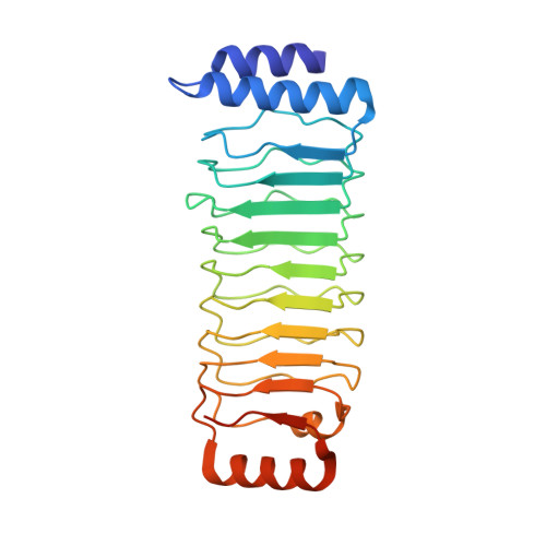
Structure of an SspH1-PKN1 Complex Reveals the Basis for Host Substrate Recognition and Mechanism of Activation for a Bacterial E3 Ubiquitin Ligase.

Keszei, A.F.Tang, X.McCormick, C.Zeqiraj, E.Rohde, J.R.Tyers, M.Sicheri, F.

(2014) Mol Cell Biol 34: 362-373

  • DOI: https://doi.org/10.1128/MCB.01360-13
  • Primary Citation of Related Structures:  
    4NKG, 4NKH

  • PubMed Abstract: 

    IpaH proteins are bacterium-specific E3 enzymes that function as type three secretion system (T3SS) effectors in Salmonella, Shigella, and other Gram-negative bacteria. IpaH enzymes recruit host substrates for ubiquitination via a leucine-rich repeat (LRR) domain, which can inhibit the catalytic domain in the absence of substrate. The basis for substrate recognition and the alleviation of autoinhibition upon substrate binding is unknown. Here, we report the X-ray structure of Salmonella SspH1 in complex with human PKN1. The LRR domain of SspH1 interacts specifically with the HR1b coiled-coil subdomain of PKN1 in a manner that sterically displaces the catalytic domain from the LRR domain, thereby activating catalytic function. SspH1 catalyzes the ubiquitination and proteasome-dependent degradation of PKN1 in cells, which attenuates androgen receptor responsiveness but not NF-κB activity. These regulatory features are conserved in other IpaH-substrate interactions. Our results explain the mechanism whereby substrate recognition and enzyme autoregulation are coupled in this class of bacterial ubiquitin ligases.


  • Organizational Affiliation

    Lunenfeld-Tanenbaum Research Institute, Mount Sinai Hospital, Toronto, Ontario, Canada.


Macromolecules
Find similar proteins by:  (by identity cutoff)  |  3D Structure
Entity ID: 1
MoleculeChains Sequence LengthOrganismDetailsImage
E3 ubiquitin-protein ligase sspH1
A, C
246Salmonella enterica subsp. enterica serovar Typhimurium str. 14028SMutation(s): 0 
Gene Names: sspH1STM14_1483
EC: 6.3.2
UniProt
Find proteins for D0ZVG2 (Salmonella typhimurium (strain 14028s / SGSC 2262))
Explore D0ZVG2 
Go to UniProtKB:  D0ZVG2
Entity Groups  
Sequence Clusters30% Identity50% Identity70% Identity90% Identity95% Identity100% Identity
UniProt GroupD0ZVG2
Sequence Annotations
Expand
  • Reference Sequence
Find similar proteins by:  (by identity cutoff)  |  3D Structure
Entity ID: 2
MoleculeChains Sequence LengthOrganismDetailsImage
Serine/threonine-protein kinase N1
B, D
82Homo sapiensMutation(s): 0 
Gene Names: PAK1PKNPKN1PRK1PRKCL1
EC: 2.7.11.13
UniProt & NIH Common Fund Data Resources
Find proteins for Q16512 (Homo sapiens)
Explore Q16512 
Go to UniProtKB:  Q16512
PHAROS:  Q16512
GTEx:  ENSG00000123143 
Entity Groups  
Sequence Clusters30% Identity50% Identity70% Identity90% Identity95% Identity100% Identity
UniProt GroupQ16512
Sequence Annotations
Expand
  • Reference Sequence
Experimental Data & Validation

Experimental Data

  • Method: X-RAY DIFFRACTION
  • Resolution: 2.90 Å
  • R-Value Free: 0.242 
  • R-Value Work: 0.209 
  • R-Value Observed: 0.210 
  • Space Group: P 32 2 1
Unit Cell:
Length ( Å )Angle ( ˚ )
a = 167.623α = 90
b = 167.623β = 90
c = 89.143γ = 120
Software Package:
Software NamePurpose
HKL-2000data collection
PHASERphasing
PHENIXrefinement
HKL-2000data reduction
HKL-2000data scaling

Structure Validation

View Full Validation Report


Entry History 

Deposition Data

Revision History  (Full details and data files)

  • Version 1.0: 2013-12-11
    Type: Initial release
  • Version 1.1: 2013-12-25
    Changes: Database references
  • Version 1.2: 2014-01-22
    Changes: Database references
  • Version 1.3: 2024-02-28
    Changes: Data collection, Database references, Derived calculations