De novo protein crystal structure determination from X-ray free-electron laser data.
Barends, T.R., Foucar, L., Botha, S., Doak, R.B., Shoeman, R.L., Nass, K., Koglin, J.E., Williams, G.J., Boutet, S., Messerschmidt, M., Schlichting, I.(2014) Nature 505: 244-247
- PubMed: 24270807
- DOI: https://doi.org/10.1038/nature12773
- Primary Citation of Related Structures:
4N5R - PubMed Abstract:
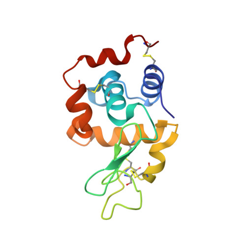
The determination of protein crystal structures is hampered by the need for macroscopic crystals. X-ray free-electron lasers (FELs) provide extremely intense pulses of femtosecond duration, which allow data collection from nanometre- to micrometre-sized crystals in a 'diffraction-before-destruction' approach. So far, all protein structure determinations carried out using FELs have been based on previous knowledge of related, known structures. Here we show that X-ray FEL data can be used for de novo protein structure determination, that is, without previous knowledge about the structure. Using the emerging technique of serial femtosecond crystallography, we performed single-wavelength anomalous scattering measurements on microcrystals of the well-established model system lysozyme, in complex with a lanthanide compound. Using Monte-Carlo integration, we obtained high-quality diffraction intensities from which experimental phases could be determined, resulting in an experimental electron density map good enough for automated building of the protein structure. This demonstrates the feasibility of determining novel protein structures using FELs. We anticipate that serial femtosecond crystallography will become an important tool for the structure determination of proteins that are difficult to crystallize, such as membrane proteins.
Organizational Affiliation:
Max-Planck Institute for Medical Research, Jahnstrasse 29, D-69120 Heidelberg, Germany.