4MZR

Crystal structure of a polypeptide p53 mutant bound to DNA


Experimental Data Snapshot

  • Method: X-RAY DIFFRACTION
  • Resolution: 2.90 Å
  • R-Value Free: 0.281 
  • R-Value Work: 0.234 
  • R-Value Observed: 0.234 

wwPDB Validation   3D Report Full Report


This is version 1.4 of the entry. See complete history


Literature

Reversal of the DNA-Binding-Induced Loop L1 Conformational Switch in an Engineered Human p53 Protein.

Emamzadah, S.Tropia, L.Vincenti, I.Falquet, B.Halazonetis, T.D.

(2014) J Mol Biol 426: 936-944

  • DOI: https://doi.org/10.1016/j.jmb.2013.12.020
  • Primary Citation of Related Structures:  
    4MZI, 4MZR

  • PubMed Abstract: 

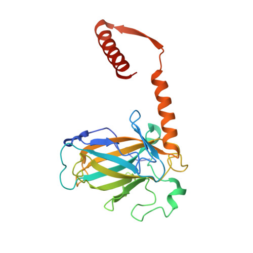
    The gene encoding the p53 tumor suppressor protein, a sequence-specific DNA binding transcription factor, is the most frequently mutated gene in human cancer. Crystal structures of homo-oligomerizing p53 polypeptides with specific DNA suggest that DNA binding is associated with a conformational switch. Specifically, in the absence of DNA, loop L1 of the p53 DNA binding domain adopts an extended conformation, whereas two p53 subunits switch to a recessed loop L1 conformation when bound to DNA as a tetramer. We previously designed a p53 protein, p53FG, with amino substitutions S121F and V122G targeting loop L1. These two substitutions enhanced the affinity of p53 for specific DNA yet, counterintuitively, decreased the residency time of p53 on DNA. Here, we confirmed these DNA binding properties of p53FG using a different method. We also determined by crystallography the structure of p53FG in its free state and bound to DNA as a tetramer. In the free state, loop L1 adopted a recessed conformation, whereas upon DNA binding, two subunits switched to the extended loop L1 conformation, resulting in a final structure that was very similar to that of wild-type p53 bound to DNA. Thus, altering the apo structure of p53 changed its DNA binding properties, even though the DNA-bound structure was not altered.


  • Organizational Affiliation

    Department of Molecular Biology, University of Geneva, 1205 Geneva, Switzerland.


Macromolecules

Find similar proteins by:  (by identity cutoff)  |  3D Structure
Entity ID: 1
MoleculeChains Sequence LengthOrganismDetailsImage
Cellular tumor antigen p53
A, B, C, D
237Homo sapiensMutation(s): 17 
Gene Names: TP53P53
UniProt & NIH Common Fund Data Resources
Find proteins for P04637 (Homo sapiens)
Explore P04637 
Go to UniProtKB:  P04637
PHAROS:  P04637
GTEx:  ENSG00000141510 
Entity Groups  
Sequence Clusters30% Identity50% Identity70% Identity90% Identity95% Identity100% Identity
UniProt GroupP04637
Sequence Annotations
Expand
  • Reference Sequence
Find similar nucleic acids by:  (by identity cutoff)  |  3D Structure
Entity ID: 2
MoleculeChains LengthOrganismImage
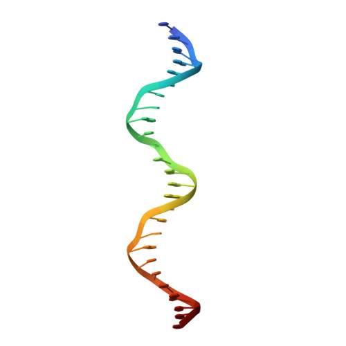
consensus DNA sense strandE [auth K]26N/A
Sequence Annotations
Expand
  • Reference Sequence
Find similar nucleic acids by:  (by identity cutoff)  |  3D Structure
Entity ID: 3
MoleculeChains LengthOrganismImage
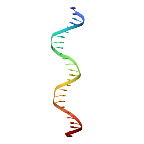
consensus DNA anti-sense strandF [auth L]26N/A
Sequence Annotations
Expand
  • Reference Sequence
Experimental Data & Validation

Experimental Data

  • Method: X-RAY DIFFRACTION
  • Resolution: 2.90 Å
  • R-Value Free: 0.281 
  • R-Value Work: 0.234 
  • R-Value Observed: 0.234 
  • Space Group: P 21 21 2
Unit Cell:
Length ( Å )Angle ( ˚ )
a = 163.298α = 90
b = 169.764β = 90
c = 55.664γ = 90
Software Package:
Software NamePurpose
MxCuBEdata collection
CNSrefinement
MOSFLMdata reduction
SCALAdata scaling
CNSphasing

Structure Validation

View Full Validation Report


Entry History 

Deposition Data

Revision History  (Full details and data files)

  • Version 1.0: 2014-01-15
    Type: Initial release
  • Version 1.1: 2014-02-12
    Changes: Database references
  • Version 1.2: 2019-02-20
    Changes: Data collection, Database references
  • Version 1.3: 2022-12-21
    Changes: Database references, Derived calculations
  • Version 1.4: 2023-09-20
    Changes: Data collection, Refinement description