4MU8

Crystal structure of an oxidized form of yeast iso-1-cytochrome c at pH 8.8


Experimental Data Snapshot

  • Method: X-RAY DIFFRACTION
  • Resolution: 1.45 Å
  • R-Value Free: 0.154 
  • R-Value Work: 0.145 
  • R-Value Observed: 0.145 

wwPDB Validation   3D Report Full Report


Ligand Structure Quality Assessment 


This is version 2.1 of the entry. See complete history


Literature

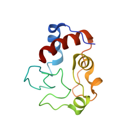
Structure of a mitochondrial cytochrome c conformer competent for peroxidase activity.

McClelland, L.J.Mou, T.C.Jeakins-Cooley, M.E.Sprang, S.R.Bowler, B.E.

(2014) Proc Natl Acad Sci U S A 111: 6648-6653

  • DOI: https://doi.org/10.1073/pnas.1323828111
  • Primary Citation of Related Structures:  
    4MU8

  • PubMed Abstract: 

    At the onset of apoptosis, the peroxidation of cardiolipin at the inner mitochondrial membrane by cytochrome c requires an open coordination site on the heme. We report a 1.45-Å resolution structure of yeast iso-1-cytochrome c with the Met80 heme ligand swung out of the heme crevice and replaced by a water molecule. This conformational change requires modest adjustments to the main chain of the heme crevice loop and is facilitated by a trimethyllysine 72-to-alanine mutation. This mutation also enhances the peroxidase activity of iso-1-cytochrome c. The structure shows a buried water channel capable of facilitating peroxide access to the active site and of moving protons produced during peroxidase activity to the protein surface. Alternate positions of the side chain of Arg38 appear to mediate opening and closing of the buried water channel. In addition, two buried water molecules can adopt alternate positions that change the network of hydrogen bonds in the buried water channel. Taken together, these observations suggest that low and high proton conductivity states may mediate peroxidase function. Comparison of yeast and mammalian cytochrome c sequences, in the context of the steric factors that permit opening of the heme crevice, suggests that higher organisms have evolved to inhibit peroxidase activity, providing a more stringent barrier to the onset of apoptosis.


  • Organizational Affiliation

    Department of Chemistry and Biochemistry, Center for Biomolecular Structure and Dynamics, and Division of Biological Sciences, University of Montana, Missoula, MT 59812.


Macromolecules
Find similar proteins by:  (by identity cutoff)  |  3D Structure
Entity ID: 1
MoleculeChains Sequence LengthOrganismDetailsImage
Cytochrome c iso-1
A, B
108Saccharomyces cerevisiae S288CMutation(s): 2 
Gene Names: CYC1YJR048WJ1653
UniProt
Find proteins for P00044 (Saccharomyces cerevisiae (strain ATCC 204508 / S288c))
Explore P00044 
Go to UniProtKB:  P00044
Entity Groups  
Sequence Clusters30% Identity50% Identity70% Identity90% Identity95% Identity100% Identity
UniProt GroupP00044
Sequence Annotations
Expand
  • Reference Sequence
Small Molecules
Ligands 4 Unique
IDChains Name / Formula / InChI Key2D Diagram3D Interactions
HEC
Query on HEC

Download Ideal Coordinates CCD File 
C [auth A],
J [auth B]
HEME C
C34 H34 Fe N4 O4
HXQIYSLZKNYNMH-LJNAALQVSA-N
SO4
Query on SO4

Download Ideal Coordinates CCD File 
D [auth A]
E [auth A]
F [auth A]
G [auth A]
I [auth B]
D [auth A],
E [auth A],
F [auth A],
G [auth A],
I [auth B],
K [auth B],
L [auth B],
M [auth B]
SULFATE ION
O4 S
QAOWNCQODCNURD-UHFFFAOYSA-L
GOL
Query on GOL

Download Ideal Coordinates CCD File 
N [auth B]GLYCEROL
C3 H8 O3
PEDCQBHIVMGVHV-UHFFFAOYSA-N
TBU
Query on TBU

Download Ideal Coordinates CCD File 
H [auth A],
O [auth B],
P [auth B]
TERTIARY-BUTYL ALCOHOL
C4 H10 O
DKGAVHZHDRPRBM-UHFFFAOYSA-N
Experimental Data & Validation

Experimental Data

  • Method: X-RAY DIFFRACTION
  • Resolution: 1.45 Å
  • R-Value Free: 0.154 
  • R-Value Work: 0.145 
  • R-Value Observed: 0.145 
  • Space Group: P 63 2 2
Unit Cell:
Length ( Å )Angle ( ˚ )
a = 93.621α = 90
b = 93.621β = 90
c = 155.014γ = 120
Software Package:
Software NamePurpose
Blu-Icedata collection
PHASESphasing
PHENIXrefinement
HKL-2000data reduction
HKL-2000data scaling

Structure Validation

View Full Validation Report


Ligand Structure Quality Assessment 


Entry History 

Deposition Data

Revision History  (Full details and data files)

  • Version 1.0: 2014-06-04
    Type: Initial release
  • Version 2.0: 2021-03-10
    Changes: Atomic model, Data collection, Database references, Derived calculations, Non-polymer description, Structure summary
  • Version 2.1: 2023-09-20
    Changes: Data collection, Database references, Refinement description