4MM3

Crystal structure of SARS-CoV papain-like protease PLpro in complex with ubiquitin aldehyde


Experimental Data Snapshot

  • Method: X-RAY DIFFRACTION
  • Resolution: 2.75 Å
  • R-Value Free: 0.279 
  • R-Value Work: 0.187 
  • R-Value Observed: 0.192 

wwPDB Validation   3D Report Full Report


This is version 1.1 of the entry. See complete history


Literature

Structural Basis for the Ubiquitin-Linkage Specificity and deISGylating activity of SARS-CoV papain-like protease.

Ratia, K.Kilianski, A.Baez-Santos, Y.M.Baker, S.C.Mesecar, A.

(2014) PLoS Pathog 10: e1004113-e1004113

  • DOI: https://doi.org/10.1371/journal.ppat.1004113
  • Primary Citation of Related Structures:  
    4MM3

  • PubMed Abstract: 

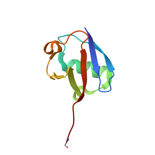
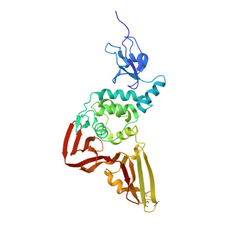
    Severe acute respiratory syndrome coronavirus (SARS-CoV) encodes a papain-like protease (PLpro) with both deubiquitinating (DUB) and deISGylating activities that are proposed to counteract the post-translational modification of signaling molecules that activate the innate immune response. Here we examine the structural basis for PLpro's ubiquitin chain and interferon stimulated gene 15 (ISG15) specificity. We present the X-ray crystal structure of PLpro in complex with ubiquitin-aldehyde and model the interaction of PLpro with other ubiquitin-chain and ISG15 substrates. We show that PLpro greatly prefers K48- to K63-linked ubiquitin chains, and ISG15-based substrates to those that are mono-ubiquitinated. We propose that PLpro's higher affinity for K48-linked ubiquitin chains and ISG15 stems from a bivalent mechanism of binding, where two ubiquitin-like domains prefer to bind in the palm domain of PLpro with the most distal ubiquitin domain interacting with a "ridge" region of the thumb domain. Mutagenesis of residues within this ridge region revealed that these mutants retain viral protease activity and the ability to catalyze hydrolysis of mono-ubiquitin. However, a select number of these mutants have a significantly reduced ability to hydrolyze the substrate ISG15-AMC, or be inhibited by K48-linked diubuiquitin. For these latter residues, we found that PLpro antagonism of the nuclear factor kappa-light-chain-enhancer of activated B-cells (NFκB) signaling pathway is abrogated. This identification of key and unique sites in PLpro required for recognition and processing of diubiquitin and ISG15 versus mono-ubiquitin and protease activity provides new insight into ubiquitin-chain and ISG15 recognition and highlights a role for PLpro DUB and deISGylase activity in antagonism of the innate immune response.


  • Organizational Affiliation

    Department of Medicinal Chemistry and Pharmacognosy, University of Illinois, Chicago, Illinois, United States of America.


Macromolecules
Find similar proteins by:  (by identity cutoff)  |  3D Structure
Entity ID: 1
MoleculeChains Sequence LengthOrganismDetailsImage
Ubiquitin76Homo sapiensMutation(s): 0 
Gene Names: UBC
UniProt & NIH Common Fund Data Resources
Find proteins for P0CG48 (Homo sapiens)
Explore P0CG48 
Go to UniProtKB:  P0CG48
PHAROS:  P0CG48
GTEx:  ENSG00000150991 
Entity Groups  
Sequence Clusters30% Identity50% Identity70% Identity90% Identity95% Identity100% Identity
UniProt GroupP0CG48
Sequence Annotations
Expand
  • Reference Sequence
Find similar proteins by:  (by identity cutoff)  |  3D Structure
Entity ID: 2
MoleculeChains Sequence LengthOrganismDetailsImage
Papain-like proteinase316Severe acute respiratory syndrome-related coronavirusMutation(s): 0 
Gene Names: nsp3
EC: 3.4.19.12 (PDB Primary Data), 3.4.22.69 (PDB Primary Data)
UniProt
Find proteins for P0C6X7 (Severe acute respiratory syndrome coronavirus)
Explore P0C6X7 
Go to UniProtKB:  P0C6X7
Entity Groups  
Sequence Clusters30% Identity50% Identity70% Identity90% Identity95% Identity100% Identity
UniProt GroupP0C6X7
Sequence Annotations
Expand
  • Reference Sequence
Small Molecules
Ligands 1 Unique
IDChains Name / Formula / InChI Key2D Diagram3D Interactions
ZN
Query on ZN

Download Ideal Coordinates CCD File 
C [auth B]ZINC ION
Zn
PTFCDOFLOPIGGS-UHFFFAOYSA-N
Modified Residues  1 Unique
IDChains TypeFormula2D DiagramParent
GLZ
Query on GLZ
A
L-PEPTIDE LINKINGC2 H5 N OGLY
Experimental Data & Validation

Experimental Data

  • Method: X-RAY DIFFRACTION
  • Resolution: 2.75 Å
  • R-Value Free: 0.279 
  • R-Value Work: 0.187 
  • R-Value Observed: 0.192 
  • Space Group: P 31 2 1
Unit Cell:
Length ( Å )Angle ( ˚ )
a = 47.147α = 90
b = 47.147β = 90
c = 332.658γ = 120
Software Package:
Software NamePurpose
SCALEPACKdata scaling
PHENIXrefinement
PDB_EXTRACTdata extraction
MAR345data collection
DENZOdata reduction
PHASERphasing

Structure Validation

View Full Validation Report


Entry History 

Deposition Data

Revision History  (Full details and data files)

  • Version 1.0: 2014-07-02
    Type: Initial release
  • Version 1.1: 2017-11-15
    Changes: Refinement description