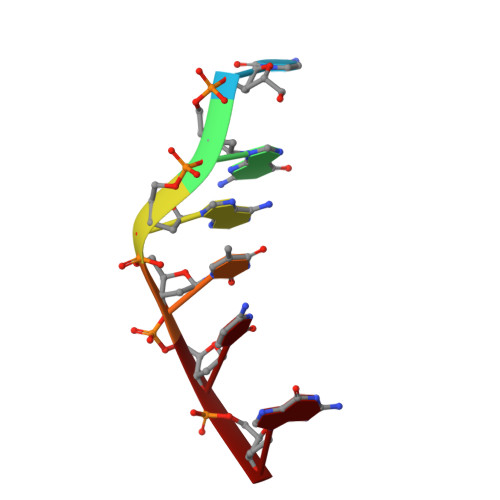
Crystal structure of the complex of DNA hexamer d(CGATCG) with Coptisine
Ferraroni, M., Bazzicalupi, C., Gratteri, P.To be published.
Experimental Data Snapshot
| Ligands 1 Unique | |||||
|---|---|---|---|---|---|
| ID | Chains | Name / Formula / InChI Key | 2D Diagram | 3D Interactions | |
| KPT Query on KPT | B [auth A] | 6,7-dihydro[1,3]dioxolo[4,5-g][1,3]dioxolo[7,8]isoquino[3,2-a]isoquinolin-5-ium C19 H14 N O4 XYHOBCMEDLZUMP-UHFFFAOYSA-N |  | ||
| Length ( Å ) | Angle ( ˚ ) |
|---|---|
| a = 26.581 | α = 90 |
| b = 26.581 | β = 90 |
| c = 77.094 | γ = 120 |
| Software Name | Purpose |
|---|---|
| XSCALE | data scaling |
| MOLREP | phasing |
| REFMAC | refinement |
| PDB_EXTRACT | data extraction |
| XDS | data reduction |
| XDS | data scaling |
RCSB PDB (citation) is hosted by
RCSB PDB is a member of the