4KWB

Structure of signal peptide peptidase A with C-termini bound in the active sites: insights into specificity, self-processing and regulation


Experimental Data Snapshot

  • Method: X-RAY DIFFRACTION
  • Resolution: 2.39 Å
  • R-Value Free: 0.240 
  • R-Value Work: 0.206 
  • R-Value Observed: 0.208 

wwPDB Validation   3D Report Full Report


This is version 1.4 of the entry. See complete history


Literature

Structure of signal peptide peptidase A with C-termini bound in the active sites: insights into specificity, self-processing, and regulation.

Nam, S.E.Paetzel, M.

(2013) Biochemistry 52: 8811-8822

  • DOI: https://doi.org/10.1021/bi4011489
  • Primary Citation of Related Structures:  
    4KWB

  • PubMed Abstract: 

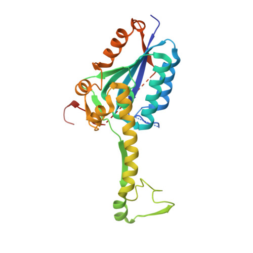
    Bacterial signal peptide peptidase A (SppA) is a membrane-bound enzyme that utilizes a serine/lysine catalytic dyad mechanism to cleave remnant signal peptides within the cellular membrane. Bacillus subtilis SppA (SppABS) oligomerizes into a homo-octameric dome-shaped complex with eight active sites, located at the interface between each protomer. In this study, we show that SppABS self-processes its own C-termini. We have determined the crystal structure of a proteolytically stable fragment of SppABSK199A that has its C-terminal peptide bound in each of the eight active sites, creating a perfect circle of peptides. Substrate specificity pockets S1, S3, and S2' are identified and accommodate C-terminal residues Tyr331, Met329, and Tyr333, respectively. Tyr331 at the P1 position is conserved among most Bacillus species. The structure reveals that the C-terminus binds within the substrate-binding grooves in an antiparallel β-sheet fashion. We show, by C-terminal truncations, that the C-terminus is not essential for oligomeric assembly. Kinetic analysis shows that a synthetic peptide corresponding to the C-terminus of SppABS competes with a fluorometric peptide substrate for the SppABS active site. A model is proposed for how the C-termini of SppA may function in the regulation of this membrane-bound self-compartmentalized protease.


  • Organizational Affiliation

    Department of Molecular Biology and Biochemistry, Simon Fraser University , South Science Building, 8888 University Drive, Burnaby, British Columbia, Canada V5A 1S6.


Macromolecules
Find similar proteins by:  (by identity cutoff)  |  3D Structure
Entity ID: 1
MoleculeChains Sequence LengthOrganismDetailsImage
Signal peptide peptidase SppA
A, B, C, D, E
A, B, C, D, E, F, G, H
273Bacillus subtilis subsp. subtilis str. 168Mutation(s): 1 
Gene Names: BSU29530CLONED SIGNAL PEPTIDE PEPTIDASE A GENE FROM RESIDUES 26 TO 335LYS199 MUTATED TO ALAsppAyteI
EC: 3.4.21
Membrane Entity: Yes 
UniProt
Find proteins for O34525 (Bacillus subtilis (strain 168))
Explore O34525 
Go to UniProtKB:  O34525
Entity Groups  
Sequence Clusters30% Identity50% Identity70% Identity90% Identity95% Identity100% Identity
UniProt GroupO34525
Sequence Annotations
Expand
  • Reference Sequence
Experimental Data & Validation

Experimental Data

  • Method: X-RAY DIFFRACTION
  • Resolution: 2.39 Å
  • R-Value Free: 0.240 
  • R-Value Work: 0.206 
  • R-Value Observed: 0.208 
  • Space Group: P 21 21 21
Unit Cell:
Length ( Å )Angle ( ˚ )
a = 87.797α = 90
b = 130.979β = 90
c = 207.099γ = 90
Software Package:
Software NamePurpose
MxDCdata collection
PHASERphasing
REFMACrefinement
HKL-2000data reduction
HKL-2000data scaling

Structure Validation

View Full Validation Report


Entry History 

Deposition Data

Revision History  (Full details and data files)

  • Version 1.0: 2013-11-27
    Type: Initial release
  • Version 1.1: 2013-12-04
    Changes: Structure summary
  • Version 1.2: 2014-02-19
    Changes: Database references
  • Version 1.3: 2017-11-15
    Changes: Refinement description
  • Version 1.4: 2023-09-20
    Changes: Data collection, Database references, Refinement description