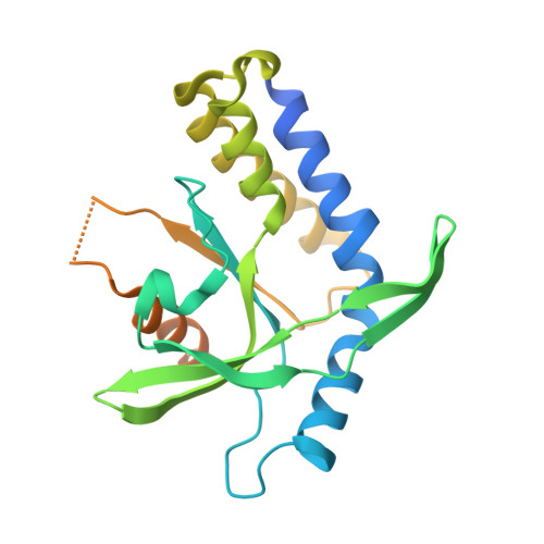
4KSY

Crystal structure of STING in complex with cGAMP


Experimental Data Snapshot

  • Method: X-RAY DIFFRACTION
  • Resolution: 1.88 Å
  • R-Value Free: 0.181 
  • R-Value Work: 0.161 
  • R-Value Observed: 0.163 

wwPDB Validation   3D Report Full Report


Ligand Structure Quality Assessment 


This is version 1.5 of the entry. See complete history


Literature

Cyclic GMP-AMP Containing Mixed Phosphodiester Linkages Is An Endogenous High-Affinity Ligand for STING.

Zhang, X.Shi, H.Wu, J.Zhang, X.Sun, L.Chen, C.Chen, Z.J.

(2013) Mol Cell 51: 226-235

  • DOI: https://doi.org/10.1016/j.molcel.2013.05.022
  • Primary Citation of Related Structures:  
    4KSY

  • PubMed Abstract: 

    The presence of microbial or self DNA in the cytoplasm of mammalian cells is a danger signal detected by the DNA sensor cyclic-GMP-AMP (cGAMP) synthase (cGAS), which catalyzes the production of cGAMP that in turn serves as a second messenger to activate innate immune responses. Here we show that endogenous cGAMP in mammalian cells contains two distinct phosphodiester linkages, one between 2'-OH of GMP and 5'-phosphate of AMP, and the other between 3'-OH of AMP and 5'-phosphate of GMP. This molecule, termed 2'3'-cGAMP, is unique in that it binds to the adaptor protein STING with a much greater affinity than cGAMP molecules containing other combinations of phosphodiester linkages. The crystal structure of STING bound to 2'3'-cGAMP revealed the structural basis of this high-affinity binding and a ligand-induced conformational change in STING that may underlie its activation.


  • Organizational Affiliation

    Department of Molecular Biology, University of Texas Southwestern Medical Center, Dallas, TX 75390-9148, USA.


Macromolecules
Find similar proteins by:  (by identity cutoff)  |  3D Structure
Entity ID: 1
MoleculeChains Sequence LengthOrganismDetailsImage
Stimulator of interferon genes protein242Homo sapiensMutation(s): 1 
Gene Names: TMEM173ERISMITASTING
UniProt & NIH Common Fund Data Resources
Find proteins for Q86WV6 (Homo sapiens)
Explore Q86WV6 
Go to UniProtKB:  Q86WV6
PHAROS:  Q86WV6
GTEx:  ENSG00000184584 
Entity Groups  
Sequence Clusters30% Identity50% Identity70% Identity90% Identity95% Identity100% Identity
UniProt GroupQ86WV6
Sequence Annotations
Expand
  • Reference Sequence
Small Molecules
Ligands 1 Unique
IDChains Name / Formula / InChI Key2D Diagram3D Interactions
1SY
Query on 1SY

Download Ideal Coordinates CCD File 
B [auth A]cGAMP
C20 H24 N10 O13 P2
XRILCFTWUCUKJR-INFSMZHSSA-N
Binding Affinity Annotations 
IDSourceBinding Affinity
1SY PDBBind:  4KSY Kd: 3.79 (nM) from 1 assay(s)
Binding MOAD:  4KSY Kd: 3.79 (nM) from 1 assay(s)
Experimental Data & Validation

Experimental Data

  • Method: X-RAY DIFFRACTION
  • Resolution: 1.88 Å
  • R-Value Free: 0.181 
  • R-Value Work: 0.161 
  • R-Value Observed: 0.163 
  • Space Group: C 1 2 1
Unit Cell:
Length ( Å )Angle ( ˚ )
a = 89.522α = 90
b = 77.927β = 96.97
c = 35.974γ = 90
Software Package:
Software NamePurpose
HKL-3000data collection
PHENIXrefinement
HKL-2000data reduction
HKL-2000data scaling

Structure Validation

View Full Validation Report


Ligand Structure Quality Assessment 


Entry History 

Deposition Data

Revision History  (Full details and data files)

  • Version 1.0: 2013-06-12
    Type: Initial release
  • Version 1.1: 2013-07-17
    Changes: Database references
  • Version 1.2: 2013-08-07
    Changes: Database references
  • Version 1.3: 2015-08-05
    Changes: Non-polymer description
  • Version 1.4: 2018-06-27
    Changes: Data collection, Database references
  • Version 1.5: 2024-02-28
    Changes: Data collection, Database references, Derived calculations, Structure summary