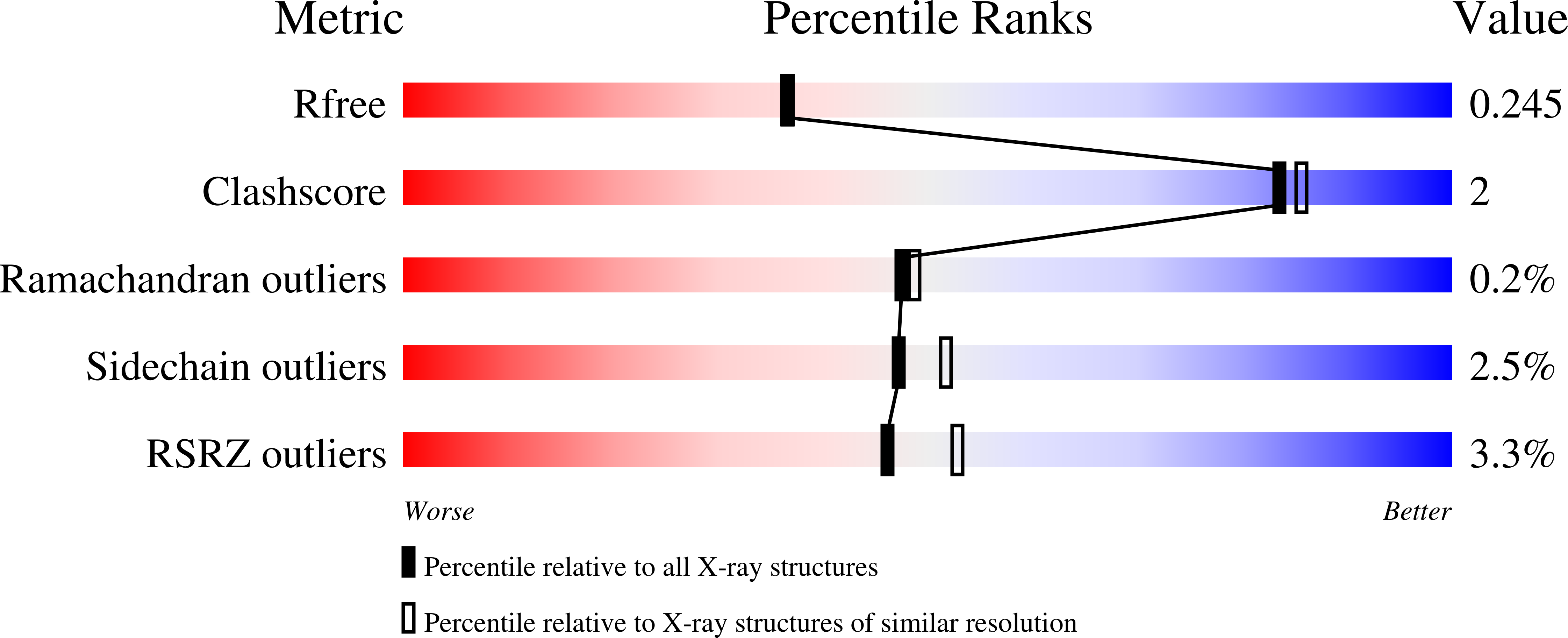
4K7R

Crystal structures of CusC review conformational changes accompanying folding and transmembrane channel formation


Experimental Data Snapshot

  • Method: X-RAY DIFFRACTION
  • Resolution: 2.09 Å
  • R-Value Free: 0.250 
  • R-Value Work: 0.214 
  • R-Value Observed: 0.216 

wwPDB Validation   3D Report Full Report


Ligand Structure Quality Assessment 


This is version 1.2 of the entry. See complete history


Literature

Crystal Structures of CusC Review Conformational Changes Accompanying Folding and Transmembrane Channel Formation.

Lei, H.T.Bolla, J.R.Bishop, N.R.Su, C.C.Yu, E.W.

(2014) J Mol Biol 426: 403-411

  • DOI: https://doi.org/10.1016/j.jmb.2013.09.042
  • Primary Citation of Related Structures:  
    4K34, 4K7K, 4K7R

  • PubMed Abstract: 

    Gram-negative bacteria, such as Escherichia coli, frequently utilize tripartite efflux complexes in the RND (resistance-nodulation-cell division) family to expel diverse toxic compounds from the cell. These complexes span both the inner and outer membranes of the bacterium via an α-helical, inner membrane transporter; a periplasmic membrane fusion protein; and a β-barrel, outer membrane channel. One such efflux system, CusCBA, is responsible for extruding biocidal Cu(I) and Ag(I) ions. To remove these toxic ions, the CusC outer membrane channel must form a β-barrel structural domain, which creates a pore and spans the entire outer membrane. We here report the crystal structures of wild-type CusC, as well as two CusC mutants, suggesting that the first N-terminal cysteine residue plays an important role in protein-membrane interactions and is critical for the insertion of this channel protein into the outer membrane. These structures provide insight into the mechanisms on CusC folding and transmembrane channel formation. It is found that the interactions between CusC and membrane may be crucial for controlling the opening and closing of this β-barrel, outer membrane channel.


  • Organizational Affiliation

    Department of Chemistry, Department of Physics & Astronomy, Iowa State University, Ames, IA 50011, USA.


Macromolecules
Find similar proteins by:  (by identity cutoff)  |  3D Structure
Entity ID: 1
MoleculeChains Sequence LengthOrganismDetailsImage
Cation efflux system protein CusC446Escherichia coli K-12Mutation(s): 0 
Gene Names: b0572cusCibeBJW0561ylcB
Membrane Entity: Yes 
UniProt
Find proteins for P77211 (Escherichia coli (strain K12))
Explore P77211 
Go to UniProtKB:  P77211
Entity Groups  
Sequence Clusters30% Identity50% Identity70% Identity90% Identity95% Identity100% Identity
UniProt GroupP77211
Sequence Annotations
Expand
  • Reference Sequence
Small Molecules
Ligands 1 Unique
IDChains Name / Formula / InChI Key2D Diagram3D Interactions
3PK
Query on 3PK

Download Ideal Coordinates CCD File 
B [auth A](2S)-1-(pentanoyloxy)propan-2-yl hexanoate
C14 H26 O4
XIGVTLHUQMMWJT-LBPRGKRZSA-N
Experimental Data & Validation

Experimental Data

  • Method: X-RAY DIFFRACTION
  • Resolution: 2.09 Å
  • R-Value Free: 0.250 
  • R-Value Work: 0.214 
  • R-Value Observed: 0.216 
  • Space Group: H 3 2
Unit Cell:
Length ( Å )Angle ( ˚ )
a = 89.026α = 90
b = 89.026β = 90
c = 474.103γ = 120
Software Package:
Software NamePurpose
SCALEPACKdata scaling
PHENIXrefinement
PDB_EXTRACTdata extraction
ADSCdata collection
DENZOdata reduction
PHASERphasing

Structure Validation

View Full Validation Report


Ligand Structure Quality Assessment 


Entry History 

Deposition Data

Revision History  (Full details and data files)

  • Version 1.0: 2013-10-16
    Type: Initial release
  • Version 1.1: 2014-02-05
    Changes: Database references
  • Version 1.2: 2024-02-28
    Changes: Data collection, Database references, Derived calculations