


SETD7 in complex with inhibitor PF-5426 and S-adenosyl-methionine
Dong, A., Wu, H., Zeng, H., El Bakkouri, M., Barsyte, D., Vedadi, M., Tatlock, J., Owen, D., Bunnage, M., Bountra, C., Arrowsmith, C.H., Edwards, A.M., Brown, P.J.To be published.
Experimental Data Snapshot
Entity ID: 1 | |||||
|---|---|---|---|---|---|
| Molecule | Chains | Sequence Length | Organism | Details | Image |
| Histone-lysine N-methyltransferase SETD7 | 264 | Homo sapiens | Mutation(s): 0 Gene Names: SETD7, KIAA1717, KMT7, SET7, SET9 EC: 2.1.1.43 | 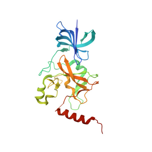 | |
UniProt & NIH Common Fund Data Resources | |||||
Find proteins for Q8WTS6 (Homo sapiens) Explore Q8WTS6 Go to UniProtKB: Q8WTS6 | |||||
PHAROS: Q8WTS6 GTEx: ENSG00000145391 | |||||
Entity Groups | |||||
| Sequence Clusters | 30% Identity50% Identity70% Identity90% Identity95% Identity100% Identity | ||||
| UniProt Group | Q8WTS6 | ||||
Sequence AnnotationsExpand | |||||
| |||||
| Ligands 3 Unique | |||||
|---|---|---|---|---|---|
| ID | Chains | Name / Formula / InChI Key | 2D Diagram | 3D Interactions | |
| 1L4 Query on 1L4 | F [auth A], FA [auth C], QA [auth D], V [auth B] | N-[(2R)-3-(3-cyanophenyl)-1-oxo-1-(pyrrolidin-1-yl)propan-2-yl]-8-fluoro-1,2,3,4-tetrahydroisoquinoline-6-sulfonamide C23 H25 F N4 O3 S HMBXWXQQVHJMOO-JOCHJYFZSA-N |  | ||
| SAM Query on SAM | E [auth A], EA [auth C], PA [auth D], U [auth B] | S-ADENOSYLMETHIONINE C15 H22 N6 O5 S MEFKEPWMEQBLKI-FCKMPRQPSA-N |  | ||
| UNX Query on UNX | AA [auth B] BA [auth B] CA [auth B] DA [auth B] G [auth A] | UNKNOWN ATOM OR ION X |  | ||
| Length ( Å ) | Angle ( ˚ ) |
|---|---|
| a = 118.048 | α = 90 |
| b = 134.482 | β = 90 |
| c = 137.255 | γ = 90 |
| Software Name | Purpose |
|---|---|
| SCALEPACK | data scaling |
| REFMAC | refinement |
| PDB_EXTRACT | data extraction |
| HKL-3000 | data reduction |
| MOLREP | phasing |
| Coot | model building |
RCSB PDB (citation) is hosted by
RCSB PDB is a member of the

