4ITU

Crystal structure of S-2-HYDROXYPROPYL COENZYME M DEHYDROGENASE (S-HPCDH) bound to S-HPC AND NADH


Experimental Data Snapshot

  • Method: X-RAY DIFFRACTION
  • Resolution: 1.60 Å
  • R-Value Free: 0.194 
  • R-Value Work: 0.164 
  • R-Value Observed: 0.166 

wwPDB Validation   3D Report Full Report


Ligand Structure Quality Assessment 


This is version 1.2 of the entry. See complete history


Literature

Crystal structures of S-HPCDH reveal determinants of stereospecificity for R- and S-hydroxypropyl-coenzyme M dehydrogenases.

Bakelar, J.W.Sliwa, D.A.Johnson, S.J.

(2013) Arch Biochem Biophys 533: 62-68

  • DOI: https://doi.org/10.1016/j.abb.2013.02.017
  • Primary Citation of Related Structures:  
    4GH5, 4ITU

  • PubMed Abstract: 

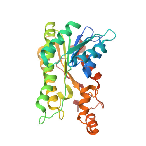
    (R)- and (S)-hydroxypropyl-coenzyme M dehydrogenases (R- and S-HPCDH) are stereospecific enzymes that are central to the metabolism of propylene and epoxide in Xanthobacter autotrophicus. The bacterium produces R- and S-HPCDH simultaneously to facilitate transformation of R- and S-enantiomers of epoxypropane to a common achiral product 2-ketopropyl-CoM (2-KPC). Both R- and S-HPCDH are highly specific for their respective substrates as each enzyme displays less than 0.5% activity with the opposite substrate isomer. In order to elucidate the structural basis for stereospecificity displayed by R- and S-HPCDH we have determined substrate bound crystal structures of S-HPCDH to 1.6Å resolution. Comparisons to the previously reported product-bound structure of R-HPCDH reveal that although the placement of catalytic residues within the active site of each enzyme is nearly identical, structural differences in the surrounding area provide each enzyme with a distinct substrate binding pocket. These structures demonstrate how chiral discrimination by R- and S-HPCDH results from alternative binding of the distal end of substrates within each substrate binding pocket.


  • Organizational Affiliation

    Department of Chemistry & Biochemistry, Utah State University, Logan, UT 84322-0300, USA.


Macromolecules
Find similar proteins by:  (by identity cutoff)  |  3D Structure
Entity ID: 1
MoleculeChains Sequence LengthOrganismDetailsImage
Short-chain dehydrogenase/reductase SDR
A, B, C, D
269Xanthobacter autotrophicus Py2Mutation(s): 0 
UniProt
Find proteins for A7IQH5 (Xanthobacter autotrophicus (strain ATCC BAA-1158 / Py2))
Explore A7IQH5 
Go to UniProtKB:  A7IQH5
Entity Groups  
Sequence Clusters30% Identity50% Identity70% Identity90% Identity95% Identity100% Identity
UniProt GroupA7IQH5
Sequence Annotations
Expand
  • Reference Sequence
Experimental Data & Validation

Experimental Data

  • Method: X-RAY DIFFRACTION
  • Resolution: 1.60 Å
  • R-Value Free: 0.194 
  • R-Value Work: 0.164 
  • R-Value Observed: 0.166 
  • Space Group: P 21 21 2
Unit Cell:
Length ( Å )Angle ( ˚ )
a = 116.048α = 90
b = 128.394β = 90
c = 58.394γ = 90
Software Package:
Software NamePurpose
HKL-3000data collection
PHASERphasing
PHENIXrefinement
HKL-2000data reduction
HKL-2000data scaling

Structure Validation

View Full Validation Report


Ligand Structure Quality Assessment 


Entry History 

Deposition Data

Revision History  (Full details and data files)

  • Version 1.0: 2013-03-27
    Type: Initial release
  • Version 1.1: 2013-04-24
    Changes: Database references
  • Version 1.2: 2023-09-20
    Changes: Data collection, Database references, Derived calculations, Refinement description