


4GG2
The crystal structure of glutamate-bound human gamma-glutamyltranspeptidase 1
- PDB DOI: https://doi.org/10.2210/pdb4GG2/pdb
- Classification: HYDROLASE
- Organism(s): Homo sapiens
- Expression System: Pichia
- Mutation(s): No
- Deposited: 2012-08-04 Released: 2013-09-25
Experimental Data Snapshot
- Method: X-RAY DIFFRACTION
- Resolution: 2.21 Å
- R-Value Free: 0.183
- R-Value Work: 0.140
- R-Value Observed: 0.142

This is version 1.4 of the entry. See complete history.

Macromolecules
Find similar proteins by:
(by identity cutoff) | 3D Structure
Entity ID: 1 | |||||
|---|---|---|---|---|---|
| Molecule | Chains | Sequence Length | Organism | Details | Image |
| Gamma-glutamyltranspeptidase 1 heavy chain | 353 | Homo sapiens | Mutation(s): 0 Gene Names: GGT, GGT1, hGGT1 EC: 2.3.2.2 (PDB Primary Data), 3.4.19.13 (PDB Primary Data), 3.4.19.14 (PDB Primary Data) | 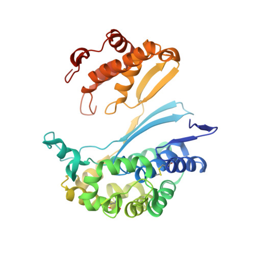 | |
UniProt & NIH Common Fund Data Resources | |||||
Find proteins for P19440 (Homo sapiens) Explore P19440 Go to UniProtKB: P19440 | |||||
PHAROS: P19440 GTEx: ENSG00000100031 | |||||
Entity Groups | |||||
| Sequence Clusters | 30% Identity50% Identity70% Identity90% Identity95% Identity100% Identity | ||||
| UniProt Group | P19440 | ||||
Sequence AnnotationsExpand | |||||
| |||||
Find similar proteins by:
(by identity cutoff) | 3D Structure
Entity ID: 2 | |||||
|---|---|---|---|---|---|
| Molecule | Chains | Sequence Length | Organism | Details | Image |
| Gamma-glutamyltranspeptidase 1 light chain | 189 | Homo sapiens | Mutation(s): 0 Gene Names: GGT, GGT1, hGGT1 EC: 2.3.2.2 (PDB Primary Data), 3.4.19.13 (PDB Primary Data), 3.4.19.14 (PDB Primary Data) | 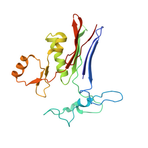 | |
UniProt & NIH Common Fund Data Resources | |||||
Find proteins for P19440 (Homo sapiens) Explore P19440 Go to UniProtKB: P19440 | |||||
PHAROS: P19440 GTEx: ENSG00000100031 | |||||
Entity Groups | |||||
| Sequence Clusters | 30% Identity50% Identity70% Identity90% Identity95% Identity100% Identity | ||||
| UniProt Group | P19440 | ||||
Sequence AnnotationsExpand | |||||
| |||||
Small Molecules
| Ligands 4 Unique | |||||
|---|---|---|---|---|---|
| ID | Chains | Name / Formula / InChI Key | 2D Diagram | 3D Interactions | |
| NAG Query on NAG | C [auth A] D [auth A] E [auth A] F [auth A] G [auth A] | 2-acetamido-2-deoxy-beta-D-glucopyranose C8 H15 N O6 OVRNDRQMDRJTHS-FMDGEEDCSA-N |  | ||
| GLU Query on GLU | N [auth B] | GLUTAMIC ACID C5 H9 N O4 WHUUTDBJXJRKMK-VKHMYHEASA-N |  | ||
| IOD Query on IOD | H [auth A] I [auth A] J [auth A] K [auth A] L [auth A] | IODIDE ION I XMBWDFGMSWQBCA-UHFFFAOYSA-M |  | ||
| CL Query on CL | S [auth B] | CHLORIDE ION Cl VEXZGXHMUGYJMC-UHFFFAOYSA-M |  | ||
Experimental Data & Validation
Experimental Data
- Method: X-RAY DIFFRACTION
- Resolution: 2.21 Å
- R-Value Free: 0.183
- R-Value Work: 0.140
- R-Value Observed: 0.142
- Space Group: C 2 2 21
Unit Cell:
| Length ( Å ) | Angle ( ˚ ) |
|---|---|
| a = 105.717 | α = 90 |
| b = 126.753 | β = 90 |
| c = 104.629 | γ = 90 |
| Software Name | Purpose |
|---|---|
| PROTEUM PLUS | data collection |
| PHENIX | model building |
| PHENIX | refinement |
| HKL-2000 | data reduction |
| HKL-2000 | data scaling |
| PHENIX | phasing |
Entry History
Deposition Data
- Released Date: 2013-09-25 Deposition Author(s): West, M.B., Chen, Y., Wickham, S., Heroux, A., Cahill, K., Hanigan, M.H., Mooers, B.H.M.
Revision History (Full details and data files)
- Version 1.0: 2013-09-25
Type: Initial release - Version 1.1: 2013-10-16
Changes: Database references - Version 1.2: 2013-11-27
Changes: Database references - Version 1.3: 2020-07-29
Type: Remediation
Reason: Carbohydrate remediation
Changes: Data collection, Derived calculations, Structure summary - Version 1.4: 2023-09-13
Changes: Data collection, Database references, Refinement description, Structure summary
