4GC9

Crystal structure of murine TFB1M in complex with SAM


Experimental Data Snapshot

  • Method: X-RAY DIFFRACTION
  • Resolution: 2.10 Å
  • R-Value Free: 0.234 
  • R-Value Work: 0.205 
  • R-Value Observed: 0.207 

wwPDB Validation   3D Report Full Report


Ligand Structure Quality Assessment 


This is version 1.3 of the entry. See complete history


Literature

Structural basis for S-adenosylmethionine binding and methyltransferase activity by mitochondrial transcription factor B1.

Guja, K.E.Venkataraman, K.Yakubovskaya, E.Shi, H.Mejia, E.Hambardjieva, E.Karzai, A.W.Garcia-Diaz, M.

(2013) Nucleic Acids Res 41: 7947-7959

  • DOI: https://doi.org/10.1093/nar/gkt547
  • Primary Citation of Related Structures:  
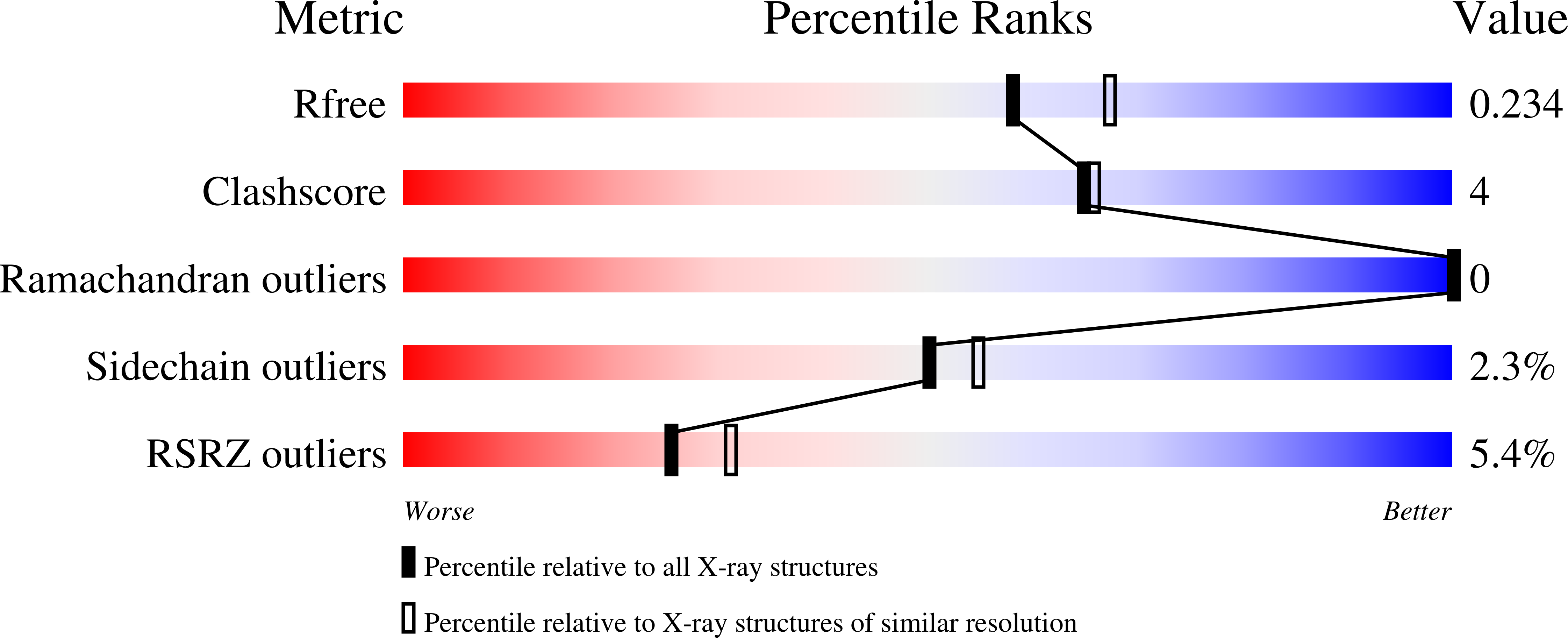
    4GC5, 4GC9

  • PubMed Abstract: 

    Eukaryotic transcription factor B (TFB) proteins are homologous to KsgA/Dim1 ribosomal RNA (rRNA) methyltransferases. The mammalian TFB1, mitochondrial (TFB1M) factor is an essential protein necessary for mitochondrial gene expression. TFB1M mediates an rRNA modification in the small ribosomal subunit and thus plays a role analogous to KsgA/Dim1 proteins. This modification has been linked to mitochondrial dysfunctions leading to maternally inherited deafness, aminoglycoside sensitivity and diabetes. Here, we present the first structural characterization of the mammalian TFB1 factor. We have solved two X-ray crystallographic structures of TFB1M with (2.1 Å) and without (2.0 Å) its cofactor S-adenosyl-L-methionine. These structures reveal that TFB1M shares a conserved methyltransferase core with other KsgA/Dim1 methyltransferases and shed light on the structural basis of S-adenosyl-L-methionine binding and methyltransferase activity. Together with mutagenesis studies, these data suggest a model for substrate binding and provide insight into the mechanism of methyl transfer, clarifying the role of this factor in an essential process for mitochondrial function.


  • Organizational Affiliation

    Department of Pharmacological Sciences, Stony Brook University, Stony Brook, NY 11794, USA, Medical Scientist Training Program, Stony Brook University Medical Center, Stony Brook, NY 11794, USA and Department of Biochemistry and Cell Biology, Stony Brook University, Stony Brook, NY 11794, USA.


Macromolecules
Find similar proteins by:  (by identity cutoff)  |  3D Structure
Entity ID: 1
MoleculeChains Sequence LengthOrganismDetailsImage
Dimethyladenosine transferase 1, mitochondrial345Mus musculusMutation(s): 0 
Gene Names: Tfb1m
EC: 2.1.1
UniProt
Find proteins for Q8JZM0 (Mus musculus)
Explore Q8JZM0 
Go to UniProtKB:  Q8JZM0
Entity Groups  
Sequence Clusters30% Identity50% Identity70% Identity90% Identity95% Identity100% Identity
UniProt GroupQ8JZM0
Sequence Annotations
Expand
  • Reference Sequence
Small Molecules
Ligands 2 Unique
IDChains Name / Formula / InChI Key2D Diagram3D Interactions
SAM
Query on SAM

Download Ideal Coordinates CCD File 
B [auth A]S-ADENOSYLMETHIONINE
C15 H22 N6 O5 S
MEFKEPWMEQBLKI-FCKMPRQPSA-N
ACT
Query on ACT

Download Ideal Coordinates CCD File 
C [auth A]ACETATE ION
C2 H3 O2
QTBSBXVTEAMEQO-UHFFFAOYSA-M
Modified Residues  1 Unique
IDChains TypeFormula2D DiagramParent
MSE
Query on MSE
A
L-PEPTIDE LINKINGC5 H11 N O2 SeMET
Experimental Data & Validation

Experimental Data

  • Method: X-RAY DIFFRACTION
  • Resolution: 2.10 Å
  • R-Value Free: 0.234 
  • R-Value Work: 0.205 
  • R-Value Observed: 0.207 
  • Space Group: I 2 2 2
Unit Cell:
Length ( Å )Angle ( ˚ )
a = 47.7α = 90
b = 101.68β = 90
c = 211.71γ = 90
Software Package:
Software NamePurpose
CBASSdata collection
PHASERphasing
PHENIXrefinement
XDSdata reduction
SCALAdata scaling

Structure Validation

View Full Validation Report


Ligand Structure Quality Assessment 


Entry History 

Deposition Data

Revision History  (Full details and data files)

  • Version 1.0: 2013-07-10
    Type: Initial release
  • Version 1.1: 2013-10-02
    Changes: Database references
  • Version 1.2: 2023-09-13
    Changes: Data collection, Database references, Derived calculations, Refinement description
  • Version 1.3: 2023-12-06
    Changes: Data collection