4FZL

High resolution structure of truncated bacteriocin syringacin M from Pseudomonas syringae pv. tomato DC3000


Experimental Data Snapshot

  • Method: X-RAY DIFFRACTION
  • Resolution: 1.46 Å
  • R-Value Free: 0.201 
  • R-Value Work: 0.152 
  • R-Value Observed: 0.154 

wwPDB Validation   3D Report Full Report


This is version 1.2 of the entry. See complete history


Literature

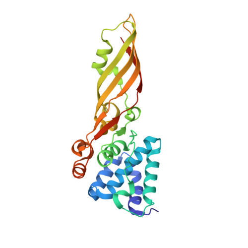
The Crystal Structure of the Lipid II-degrading Bacteriocin Syringacin M Suggests Unexpected Evolutionary Relationships between Colicin M-like Bacteriocins.

Grinter, R.Roszak, A.W.Cogdell, R.J.Milner, J.J.Walker, D.

(2012) J Biol Chem 287: 38876-38888

  • DOI: https://doi.org/10.1074/jbc.M112.400150
  • Primary Citation of Related Structures:  
    4FZL, 4FZM, 4FZN

  • PubMed Abstract: 

    Colicin-like bacteriocins show potential as next generation antibiotics with clinical and agricultural applications. Key to these potential applications is their high potency and species specificity that enables a single pathogenic species to be targeted with minimal disturbance of the wider microbial community. Here we present the structure and function of the colicin M-like bacteriocin, syringacin M from Pseudomonas syringae pv. tomato DC3000. Syringacin M kills susceptible cells through a highly specific phosphatase activity that targets lipid II, ultimately inhibiting peptidoglycan synthesis. Comparison of the structures of syringacin M and colicin M reveals that, in addition to the expected similarity between the homologous C-terminal catalytic domains, the receptor binding domains of these proteins, which share no discernible sequence homology, share a striking structural similarity. This indicates that the generation of the novel receptor binding and species specificities of these bacteriocins has been driven by diversifying selection rather than diversifying recombination as suggested previously. Additionally, the structure of syringacin M reveals the presence of an active site calcium ion that is coordinated by a conserved aspartic acid side chain and is essential for catalytic activity. We show that mutation of this residue to alanine inactivates syringacin M and that the metal ion is absent from the structure of the mutant protein. Consistent with the presence of Ca(2+) in the active site, we show that syringacin M activity is supported by Ca(2+), along with Mg(2+) and Mn(2+), and the protein is catalytically inactive in the absence of these ions.


  • Organizational Affiliation

    Institute of Infection, Immunity, and Inflammation, School of Life Sciences, College of Medical, Veterinary, and Life Sciences, University of Glasgow, Glasgow G12 8TA, Scotland, United Kingdom.


Macromolecules
Find similar proteins by:  (by identity cutoff)  |  3D Structure
Entity ID: 1
MoleculeChains Sequence LengthOrganismDetailsImage
Bacteriocin
A, B
247Pseudomonas syringae pv. tomato str. DC3000Mutation(s): 0 
Gene Names: PSPTO_0572
UniProt
Find proteins for Q88A25 (Pseudomonas syringae pv. tomato (strain ATCC BAA-871 / DC3000))
Explore Q88A25 
Go to UniProtKB:  Q88A25
Entity Groups  
Sequence Clusters30% Identity50% Identity70% Identity90% Identity95% Identity100% Identity
UniProt GroupQ88A25
Sequence Annotations
Expand
  • Reference Sequence
Small Molecules
Ligands 4 Unique
IDChains Name / Formula / InChI Key2D Diagram3D Interactions
EDO
Query on EDO

Download Ideal Coordinates CCD File 
D [auth A]
E [auth A]
F [auth A]
G [auth A]
H [auth A]
D [auth A],
E [auth A],
F [auth A],
G [auth A],
H [auth A],
I [auth A],
K [auth A],
N [auth B],
O [auth B],
P [auth B],
Q [auth B],
R [auth B],
S [auth B]
1,2-ETHANEDIOL
C2 H6 O2
LYCAIKOWRPUZTN-UHFFFAOYSA-N
CA
Query on CA

Download Ideal Coordinates CCD File 
C [auth A],
L [auth B]
CALCIUM ION
Ca
BHPQYMZQTOCNFJ-UHFFFAOYSA-N
CL
Query on CL

Download Ideal Coordinates CCD File 
J [auth A],
T [auth B]
CHLORIDE ION
Cl
VEXZGXHMUGYJMC-UHFFFAOYSA-M
MG
Query on MG

Download Ideal Coordinates CCD File 
M [auth B]MAGNESIUM ION
Mg
JLVVSXFLKOJNIY-UHFFFAOYSA-N
Experimental Data & Validation

Experimental Data

  • Method: X-RAY DIFFRACTION
  • Resolution: 1.46 Å
  • R-Value Free: 0.201 
  • R-Value Work: 0.152 
  • R-Value Observed: 0.154 
  • Space Group: P 1 21 1
Unit Cell:
Length ( Å )Angle ( ˚ )
a = 55.81α = 90
b = 96.09β = 114.17
c = 64.27γ = 90
Software Package:
Software NamePurpose
EDNAdata collection
PHASERphasing
REFMACrefinement
XDSdata reduction
SCALAdata scaling

Structure Validation

View Full Validation Report


Entry History 

Deposition Data

Revision History  (Full details and data files)

  • Version 1.0: 2012-10-03
    Type: Initial release
  • Version 1.1: 2012-11-28
    Changes: Database references
  • Version 1.2: 2024-02-28
    Changes: Data collection, Database references, Derived calculations