


Inhibitor Sites of Unequal Affinity Linked by Binding Synergism in Mutant Forms of Recombinant Human Hexokinase Type-I
Shen, L., Gao, Y., Honzatko, R.B.To be published.
Experimental Data Snapshot
Entity ID: 1 | |||||
|---|---|---|---|---|---|
| Molecule | Chains | Sequence Length | Organism | Details | Image |
| Hexokinase-1 | 917 | Homo sapiens | Mutation(s): 0 Gene Names: HK1 EC: 2.7.1.1 | 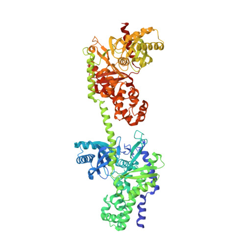 | |
UniProt & NIH Common Fund Data Resources | |||||
Find proteins for P19367 (Homo sapiens) Explore P19367 Go to UniProtKB: P19367 | |||||
PHAROS: P19367 GTEx: ENSG00000156515 | |||||
Entity Groups | |||||
| Sequence Clusters | 30% Identity50% Identity70% Identity90% Identity95% Identity100% Identity | ||||
| UniProt Group | P19367 | ||||
Sequence AnnotationsExpand | |||||
| |||||
| Ligands 4 Unique | |||||
|---|---|---|---|---|---|
| ID | Chains | Name / Formula / InChI Key | 2D Diagram | 3D Interactions | |
| M6D Query on M6D | D [auth A], F [auth A], K [auth B], M [auth B] | 6-O-phosphono-beta-D-mannopyranose C6 H13 O9 P NBSCHQHZLSJFNQ-RWOPYEJCSA-N |  | ||
| CIT Query on CIT | I [auth A], P [auth B] | CITRIC ACID C6 H8 O7 KRKNYBCHXYNGOX-UHFFFAOYSA-N |  | ||
| BGC Query on BGC | C [auth A], E [auth A], J [auth B], L [auth B] | beta-D-glucopyranose C6 H12 O6 WQZGKKKJIJFFOK-VFUOTHLCSA-N |  | ||
| NA Query on NA | G [auth A], H [auth A], N [auth B], O [auth B] | SODIUM ION Na FKNQFGJONOIPTF-UHFFFAOYSA-N |  | ||
| Length ( Å ) | Angle ( ˚ ) |
|---|---|
| a = 82.44 | α = 90 |
| b = 122.09 | β = 92.65 |
| c = 118.95 | γ = 90 |
| Software Name | Purpose |
|---|---|
| d*TREK | data reduction |
| SCALEPACK | data scaling |
| REFMAC | refinement |
| PDB_EXTRACT | data extraction |
| AMoRE | phasing |
RCSB PDB (citation) is hosted by
RCSB PDB is a member of the

