Structural, Biochemical, and Functional Characterization of the Cyclic Nucleotide Binding Homology Domain from the Mouse EAG1 Potassium Channel.
Marques-Carvalho, M.J., Sahoo, N., Muskett, F.W., Vieira-Pires, R.S., Gabant, G., Cadene, M., Schonherr, R., Morais-Cabral, J.H.(2012) J Mol Biol 423: 34-46
- PubMed: 22732247
- DOI: https://doi.org/10.1016/j.jmb.2012.06.025
- Primary Citation of Related Structures:
4F8A - PubMed Abstract:
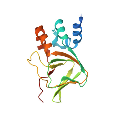
KCNH channels are voltage-gated potassium channels with important physiological functions. In these channels, a C-terminal cytoplasmic region, known as the cyclic nucleotide binding homology (CNB-homology) domain displays strong sequence similarity to cyclic nucleotide binding (CNB) domains. However, the isolated domain does not bind cyclic nucleotides. Here, we report the X-ray structure of the CNB-homology domain from the mouse EAG1 channel. Through comparison with the recently determined structure of the CNB-homology domain from the zebrafish ELK (eag-like K(+)) channel and the CNB domains from the MlotiK1 and HCN (hyperpolarization-activated cyclic nucleotide-gated) potassium channels, we establish the structural features of CNB-homology domains that explain the low affinity for cyclic nucleotides. Our structure establishes that the "self-liganded" conformation, where two residues of the C-terminus of the domain are bound in an equivalent position to cyclic nucleotides in CNB domains, is a conserved feature of CNB-homology domains. Importantly, we provide biochemical evidence that suggests that there is also an unliganded conformation where the C-terminus of the domain peels away from its bound position. A functional characterization of this unliganded conformation reveals a role of the CNB-homology domain in channel gating.
Organizational Affiliation:
Instituto de Biologia Molecular e Celular, Universidade do Porto, Rua do Campo Alegre 823, 4150-180 Porto, Portugal.