4BXK

Crystal structure of the Angiotensin-1 converting enzyme N-domain in complex with a domain-specific inhibitor


Experimental Data Snapshot

  • Method: X-RAY DIFFRACTION
  • Resolution: 2.20 Å
  • R-Value Free: 0.220 
  • R-Value Work: 0.187 
  • R-Value Observed: 0.188 

wwPDB Validation   3D Report Full Report


Ligand Structure Quality Assessment 


This is version 2.1 of the entry. See complete history


Literature

Fragment-Based Design for the Development of N-Domain Selective Angiotensin-1 Converting Enzyme Inhibitors

Douglas, R.G.Sharma, R.K.Masuyer, G.Lubbe, L.Zamora, I.Acharya, K.R.Chibale, K.Sturrock, E.D.

(2014) Clin Sci (Lond) 126: 305

  • DOI: https://doi.org/10.1042/CS20130403
  • Primary Citation of Related Structures:  
    4BXK

  • PubMed Abstract: 

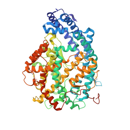
    ACE (angiotensin-1-converting enzyme) is a zinc metallopeptidase that plays a prominent role in blood pressure regulation and electrolyte homeostasis. ACE consists of two homologous domains that despite similarities of sequence and topology display differences in substrate processing and inhibitor binding. The design of inhibitors that selectively inhibit the N-domain (N-selective) could be useful in treating conditions of tissue injury and fibrosis due to build-up of N-domain-specific substrate Ac-SDKP (N-acetyl-Ser-Asp-Lys-Pro). Using a receptor-based SHOP (scaffold hopping) approach with N-selective inhibitor RXP407, a shortlist of scaffolds that consisted of modified RXP407 backbones with novel chemotypes was generated. These scaffolds were selected on the basis of enhanced predicted interaction energies with N-domain residues that differed from their C-domain counterparts. One scaffold was synthesized and inhibitory binding tested using a fluorogenic ACE assay. A molecule incorporating a tetrazole moiety in the P2 position (compound 33RE) displayed potent inhibition (K(i)=11.21±0.74 nM) and was 927-fold more selective for the N-domain than the C-domain. A crystal structure of compound 33RE in complex with the N-domain revealed its mode of binding through aromatic stacking with His388 and a direct hydrogen bond with the hydroxy group of the N-domain specific Tyr369. This work further elucidates the molecular basis for N-domain-selective inhibition and assists in the design of novel N-selective ACE inhibitors that could be employed in treatment of fibrosis disorders.


  • Organizational Affiliation

    *Institute of Infectious Disease and Molecular Medicine, and Division of Medical Biochemistry, University of Cape Town, Observatory, Cape Town 7935, South Africa.


Macromolecules
Find similar proteins by:  (by identity cutoff)  |  3D Structure
Entity ID: 1
MoleculeChains Sequence LengthOrganismDetailsImage
ANGIOTENSIN-CONVERTING ENZYME
A, B
629Homo sapiensMutation(s): 8 
EC: 3.2.1 (PDB Primary Data), 3.4.15.1 (PDB Primary Data)
UniProt & NIH Common Fund Data Resources
Find proteins for P12821 (Homo sapiens)
Explore P12821 
Go to UniProtKB:  P12821
PHAROS:  P12821
GTEx:  ENSG00000159640 
Entity Groups  
Sequence Clusters30% Identity50% Identity70% Identity90% Identity95% Identity100% Identity
UniProt GroupP12821
Sequence Annotations
Expand
  • Reference Sequence
Oligosaccharides

Help

Entity ID: 2
MoleculeChains Length2D Diagram Glycosylation3D Interactions
alpha-L-fucopyranose-(1-6)-2-acetamido-2-deoxy-beta-D-glucopyranose
C
2N-Glycosylation
Glycosylation Resources
GlyTouCan:  G86851RC
GlyCosmos:  G86851RC
GlyGen:  G86851RC
Entity ID: 3
MoleculeChains Length2D Diagram Glycosylation3D Interactions
2-acetamido-2-deoxy-beta-D-glucopyranose-(1-4)-2-acetamido-2-deoxy-beta-D-glucopyranose
D, F
2N-Glycosylation
Glycosylation Resources
GlyTouCan:  G42666HT
GlyCosmos:  G42666HT
GlyGen:  G42666HT
Entity ID: 4
MoleculeChains Length2D Diagram Glycosylation3D Interactions
beta-D-mannopyranose-(1-4)-2-acetamido-2-deoxy-beta-D-glucopyranose-(1-4)-[alpha-L-fucopyranose-(1-6)]2-acetamido-2-deoxy-beta-D-glucopyranose
E
4N-Glycosylation
Glycosylation Resources
GlyTouCan:  G32152BH
GlyCosmos:  G32152BH
GlyGen:  G32152BH
Entity ID: 5
MoleculeChains Length2D Diagram Glycosylation3D Interactions
beta-D-mannopyranose-(1-4)-2-acetamido-2-deoxy-beta-D-glucopyranose-(1-4)-2-acetamido-2-deoxy-beta-D-glucopyranose
G
3N-Glycosylation
Glycosylation Resources
GlyTouCan:  G15407YE
GlyCosmos:  G15407YE
GlyGen:  G15407YE
Small Molecules
Ligands 7 Unique
IDChains Name / Formula / InChI Key2D Diagram3D Interactions
1IU
Query on 1IU

Download Ideal Coordinates CCD File 
L [auth A],
S [auth B]
[3-[[(2S)-1-azanyl-1-oxidanylidene-propan-2-yl]amino]-2-methyl-3-oxidanylidene-propyl]-[(1R)-1-[[(2R)-2-azanyl-3-(1H-1,2,3,4-tetrazol-5-yl)propanoyl]amino]-2-phenyl-ethyl]phosphinic acid
C19 H29 N8 O5 P
RAFOALGGQUIRNB-FAXLKDOZSA-N
P6G
Query on P6G

Download Ideal Coordinates CCD File 
K [auth A],
Q [auth B]
HEXAETHYLENE GLYCOL
C12 H26 O7
IIRDTKBZINWQAW-UHFFFAOYSA-N
NAG
Query on NAG

Download Ideal Coordinates CCD File 
T [auth B]2-acetamido-2-deoxy-beta-D-glucopyranose
C8 H15 N O6
OVRNDRQMDRJTHS-FMDGEEDCSA-N
PG4
Query on PG4

Download Ideal Coordinates CCD File 
P [auth B]TETRAETHYLENE GLYCOL
C8 H18 O5
UWHCKJMYHZGTIT-UHFFFAOYSA-N
PEG
Query on PEG

Download Ideal Coordinates CCD File 
H [auth A],
I [auth A],
J [auth A],
O [auth B],
R [auth B]
DI(HYDROXYETHYL)ETHER
C4 H10 O3
MTHSVFCYNBDYFN-UHFFFAOYSA-N
ZN
Query on ZN

Download Ideal Coordinates CCD File 
M [auth A],
U [auth B]
ZINC ION
Zn
PTFCDOFLOPIGGS-UHFFFAOYSA-N
CL
Query on CL

Download Ideal Coordinates CCD File 
N [auth A],
V [auth B]
CHLORIDE ION
Cl
VEXZGXHMUGYJMC-UHFFFAOYSA-M
Binding Affinity Annotations 
IDSourceBinding Affinity
1IU PDBBind:  4BXK Ki: 11.21 (nM) from 1 assay(s)
Binding MOAD:  4BXK Ki: 11.21 (nM) from 1 assay(s)
Experimental Data & Validation

Experimental Data

  • Method: X-RAY DIFFRACTION
  • Resolution: 2.20 Å
  • R-Value Free: 0.220 
  • R-Value Work: 0.187 
  • R-Value Observed: 0.188 
  • Space Group: P 1
Unit Cell:
Length ( Å )Angle ( ˚ )
a = 73.142α = 88.43
b = 77.255β = 64.28
c = 82.866γ = 75.29
Software Package:
Software NamePurpose
REFMACrefinement
XDSdata reduction
SCALAdata scaling
PHASERphasing

Structure Validation

View Full Validation Report


Ligand Structure Quality Assessment 


Entry History 

Deposition Data

Revision History  (Full details and data files)

  • Version 1.0: 2013-09-18
    Type: Initial release
  • Version 1.1: 2013-09-25
    Changes: Database references, Derived calculations
  • Version 1.2: 2013-11-06
    Changes: Database references
  • Version 1.3: 2013-11-13
    Changes: Atomic model, Derived calculations, Other, Refinement description
  • Version 2.0: 2020-07-29
    Type: Remediation
    Reason: Carbohydrate remediation
    Changes: Advisory, Atomic model, Data collection, Derived calculations, Other, Structure summary
  • Version 2.1: 2023-12-20
    Changes: Data collection, Database references, Refinement description, Structure summary