4B1U

Structure of the Phactr1 RPEL domain and RPEL motif directed assemblies with G-actin reveal the molecular basis for actin binding cooperativity.


Experimental Data Snapshot

  • Method: X-RAY DIFFRACTION
  • Resolution: 2.00 Å
  • R-Value Free: 0.236 
  • R-Value Work: 0.195 
  • R-Value Observed: 0.197 

wwPDB Validation   3D Report Full Report


This is version 1.4 of the entry. See complete history


Literature

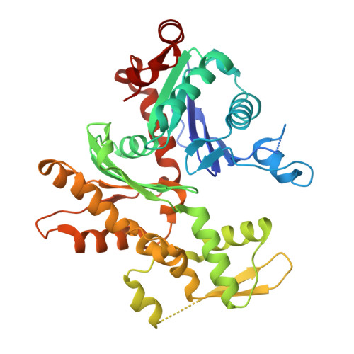
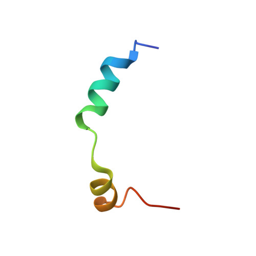
Structures of the Phactr1 RPEL domain and RPEL motif complexes with G-actin reveal the molecular basis for actin binding cooperativity.

Mouilleron, S.Wiezlak, M.O'Reilly, N.Treisman, R.McDonald, N.Q.

(2012) Structure 20: 1960-1970

  • DOI: https://doi.org/10.1016/j.str.2012.08.031
  • Primary Citation of Related Structures:  
    4B1U, 4B1V, 4B1W, 4B1X, 4B1Y, 4B1Z

  • PubMed Abstract: 

    The Phactr family of PP1-binding proteins and the myocardin-related transcription factor family of transcriptional coactivators contain regulatory domains comprising three copies of the RPEL motif, a G-actin binding element. We report the structure of a Phactr1 G-actin⋅RPEL domain complex. Three G-actins surround the crank-shaped RPEL domain forming a closed helical assembly. Their spatial relationship is identical to the RPEL-actins within the pentavalent MRTF G-actin⋅RPEL domain complex, suggesting that conserved cooperative interactions between actin⋅RPEL units organize the assembly. In the trivalent Phactr1 complex, each G-actin⋅RPEL unit makes secondary contacts with its downstream actin involving distinct RPEL residues. Similar secondary contacts are seen in G-actin⋅RPEL peptide crystals. Loss-of-secondary-contact mutations destabilize the Phactr1 G-actin⋅RPEL assembly. Furthermore, actin-mediated inhibition of Phactr1 nuclear import requires secondary contact residues in the Phactr1 N-terminal RPEL-N motif, suggesting that it involves interaction of RPEL-N with the C-terminal assembly. Secondary actin contacts by actin-bound RPEL motifs thus govern formation of multivalent actin⋅RPEL assemblies.


  • Organizational Affiliation

    Structural Biology, CRUK London Research Institute, 44 Lincoln's Inn Fields, London WC2A 3LY, UK.


Macromolecules
Find similar proteins by:  (by identity cutoff)  |  3D Structure
Entity ID: 1
MoleculeChains Sequence LengthOrganismDetailsImage
ACTIN, ALPHA SKELETAL MUSCLEA [auth B]376Oryctolagus cuniculusMutation(s): 0 
UniProt
Find proteins for P68134 (Mus musculus)
Explore P68134 
Go to UniProtKB:  P68134
Entity Groups  
Sequence Clusters30% Identity50% Identity70% Identity90% Identity95% Identity100% Identity
UniProt GroupP68134
Sequence Annotations
Expand
  • Reference Sequence
Find similar proteins by:  (by identity cutoff)  |  3D Structure
Entity ID: 2
MoleculeChains Sequence LengthOrganismDetailsImage
PHOSPHATASE AND ACTIN REGULATOR 1B [auth M]32Oryctolagus cuniculusMutation(s): 0 
UniProt & NIH Common Fund Data Resources
Find proteins for Q2M3X8 (Mus musculus)
Explore Q2M3X8 
Go to UniProtKB:  Q2M3X8
IMPC:  MGI:2659021
Entity Groups  
Sequence Clusters30% Identity50% Identity70% Identity90% Identity95% Identity100% Identity
UniProt GroupQ2M3X8
Sequence Annotations
Expand
  • Reference Sequence
Small Molecules
Ligands 5 Unique
IDChains Name / Formula / InChI Key2D Diagram3D Interactions
ATP
Query on ATP

Download Ideal Coordinates CCD File 
D [auth B]ADENOSINE-5'-TRIPHOSPHATE
C10 H16 N5 O13 P3
ZKHQWZAMYRWXGA-KQYNXXCUSA-N
LAB
Query on LAB

Download Ideal Coordinates CCD File 
C [auth B]LATRUNCULIN B
C20 H29 N O5 S
NSHPHXHGRHSMIK-JRIKCGFMSA-N
TRS
Query on TRS

Download Ideal Coordinates CCD File 
F [auth B]2-AMINO-2-HYDROXYMETHYL-PROPANE-1,3-DIOL
C4 H12 N O3
LENZDBCJOHFCAS-UHFFFAOYSA-O
CA
Query on CA

Download Ideal Coordinates CCD File 
G [auth M]CALCIUM ION
Ca
BHPQYMZQTOCNFJ-UHFFFAOYSA-N
MG
Query on MG

Download Ideal Coordinates CCD File 
E [auth B]MAGNESIUM ION
Mg
JLVVSXFLKOJNIY-UHFFFAOYSA-N
Experimental Data & Validation

Experimental Data

  • Method: X-RAY DIFFRACTION
  • Resolution: 2.00 Å
  • R-Value Free: 0.236 
  • R-Value Work: 0.195 
  • R-Value Observed: 0.197 
  • Space Group: P 32 2 1
Unit Cell:
Length ( Å )Angle ( ˚ )
a = 77.6α = 90
b = 77.6β = 90
c = 128.23γ = 120
Software Package:
Software NamePurpose
PHENIXrefinement
PHASERphasing

Structure Validation

View Full Validation Report


Entry History 

Deposition Data

Revision History  (Full details and data files)

  • Version 1.0: 2013-07-31
    Type: Initial release
  • Version 1.1: 2014-10-22
    Changes: Database references
  • Version 1.2: 2018-02-28
    Changes: Database references
  • Version 1.3: 2018-05-02
    Changes: Data collection
  • Version 1.4: 2023-12-20
    Changes: Data collection, Database references, Derived calculations, Other, Refinement description