4AT7

Crystal structure of the NF90-NF45 dimerisation domain complex


Experimental Data Snapshot

  • Method: X-RAY DIFFRACTION
  • Resolution: 1.90 Å
  • R-Value Free: 0.210 
  • R-Value Work: 0.185 
  • R-Value Observed: 0.186 

wwPDB Validation   3D Report Full Report


This is version 1.2 of the entry. See complete history


Literature

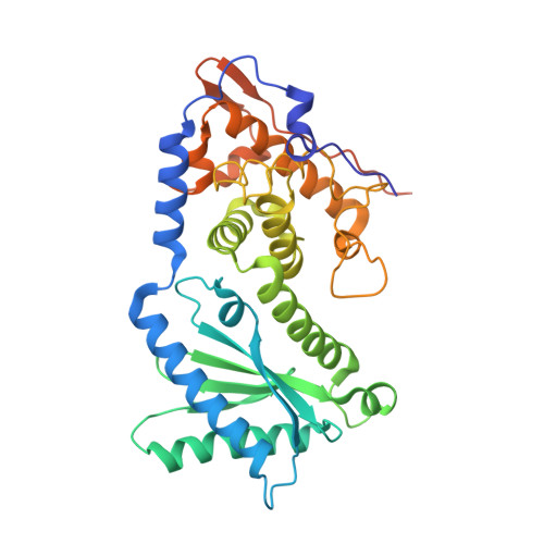
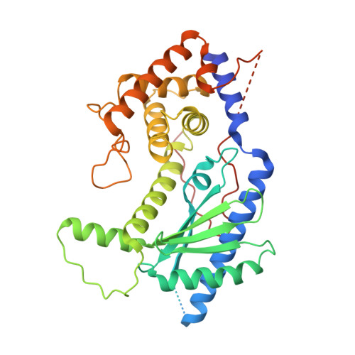
NF45 Dimerizes with NF90, Zfr and Spnr Via a Conserved Domain that Has a Nucleotidyltransferase Fold.

Wolkowicz, U.M.Cook, A.G.

(2012) Nucleic Acids Res 40: 9356

  • DOI: https://doi.org/10.1093/nar/gks696
  • Primary Citation of Related Structures:  
    4AT7, 4AT8, 4AT9, 4ATB

  • PubMed Abstract: 

    Nuclear factors NF90 and NF45 form a complex involved in a variety of cellular processes and are thought to affect gene expression both at the transcriptional and translational level. In addition, this complex affects the replication of several viruses through direct interactions with viral RNA. NF90 and NF45 dimerize through their common 'DZF' domain (domain associated with zinc fingers). NF90 has additional double-stranded RNA-binding domains that likely mediate its association with target RNAs. We present the crystal structure of the NF90/NF45 dimerization complex at 1.9-Å resolution. The DZF domain shows structural similarity to the template-free nucleotidyltransferase family of RNA modifying enzymes. However, both NF90 and NF45 have lost critical catalytic residues during evolution and are therefore not functional enzymes. Residues on NF90 that make up its interface with NF45 are conserved in two related proteins, spermatid perinuclear RNA-binding protein (SPNR) and zinc-finger RNA-binding protein (Zfr). Using a co-immunoprecipitation assay and site-specific mutants, we demonstrate that NF45 is also able to recognize SPNR and Zfr through the same binding interface, revealing that NF45 is able to form a variety of cellular complexes with other DZF-domain proteins.


  • Organizational Affiliation

    Wellcome Trust Centre for Cell Biology, University of Edinburgh, Michael Swann Building, Edinburgh, Midlothian EH9 3JR, UK.


Macromolecules
Find similar proteins by:  (by identity cutoff)  |  3D Structure
Entity ID: 1
MoleculeChains Sequence LengthOrganismDetailsImage
INTERLEUKIN ENHANCER-BINDING FACTOR 2364Mus musculusMutation(s): 0 
UniProt
Find proteins for Q9CXY6 (Mus musculus)
Explore Q9CXY6 
Go to UniProtKB:  Q9CXY6
Entity Groups  
Sequence Clusters30% Identity50% Identity70% Identity90% Identity95% Identity100% Identity
UniProt GroupQ9CXY6
Sequence Annotations
Expand
  • Reference Sequence
Find similar proteins by:  (by identity cutoff)  |  3D Structure
Entity ID: 2
MoleculeChains Sequence LengthOrganismDetailsImage
INTERLEUKIN ENHANCER-BINDING FACTOR 3383Mus musculusMutation(s): 0 
UniProt
Find proteins for Q9Z1X4 (Mus musculus)
Explore Q9Z1X4 
Go to UniProtKB:  Q9Z1X4
Entity Groups  
Sequence Clusters30% Identity50% Identity70% Identity90% Identity95% Identity100% Identity
UniProt GroupQ9Z1X4
Sequence Annotations
Expand
  • Reference Sequence
Experimental Data & Validation

Experimental Data

  • Method: X-RAY DIFFRACTION
  • Resolution: 1.90 Å
  • R-Value Free: 0.210 
  • R-Value Work: 0.185 
  • R-Value Observed: 0.186 
  • Space Group: I 2 2 2
Unit Cell:
Length ( Å )Angle ( ˚ )
a = 82.9α = 90
b = 131.3β = 90
c = 176γ = 90
Software Package:
Software NamePurpose
PHENIXrefinement
XDSdata reduction
XSCALEdata scaling
PHASERphasing

Structure Validation

View Full Validation Report


Entry History 

Deposition Data

Revision History  (Full details and data files)

  • Version 1.0: 2012-08-01
    Type: Initial release
  • Version 1.1: 2012-10-31
    Changes: Database references
  • Version 1.2: 2013-04-17
    Changes: Database references