3VMK

3-isopropylmalate dehydrogenase from Shewanella benthica DB21 MT-2


Experimental Data Snapshot

  • Method: X-RAY DIFFRACTION
  • Resolution: 1.48 Å
  • R-Value Free: 0.232 
  • R-Value Work: 0.193 
  • R-Value Observed: 0.195 

wwPDB Validation   3D Report Full Report


Ligand Structure Quality Assessment 


This is version 1.3 of the entry. See complete history


Literature

Structural analysis of 3-isopropylmalate dehydrogenase from the obligate piezophile Shewanella benthica DB21MT-2 and the nonpiezophile Shewanella oneidensis MR-1

Nagae, T.Kato, C.Watanabe, N.

(2012) Acta Crystallogr Sect F Struct Biol Cryst Commun 68: 265-268

  • DOI: https://doi.org/10.1107/S1744309112001443
  • Primary Citation of Related Structures:  
    3VMJ, 3VMK

  • PubMed Abstract: 

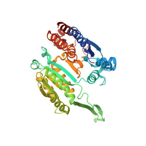
    Organisms living in deep seas such as the Mariana Trench must be adapted to the extremely high pressure environment. For example, the 3-isopropylmalate dehydrogenase from the obligate piezophile Shewanella benthica DB21MT-2 (SbIPMDH) remains active in extreme conditions under which that from the land bacterium S. oneidensis MR-1 (SoIPMDH) becomes inactivated. In order to unravel the differences between these two IPMDHs, their structures were determined at ~1.5 Å resolution. Comparison of the structures of the two enzymes shows that SbIPMDH is in a more open form and has a larger internal cavity volume than SoIPMDH at atmospheric pressure. This loosely packed structure of SbIPMDH could help it to avoid pressure-induced distortion of the native structure and to remain active at higher pressures than SoIPMDH.


  • Organizational Affiliation

    Department of Biotechnology, Graduate School of Engineering, Nagoya University, Japan.


Macromolecules
Find similar proteins by:  (by identity cutoff)  |  3D Structure
Entity ID: 1
MoleculeChains Sequence LengthOrganismDetailsImage
3-isopropylmalate dehydrogenase
A, B
375Shewanella benthicaMutation(s): 0 
Gene Names: leuB
EC: 1.1.1.85
UniProt
Find proteins for D2YZL2 (Shewanella benthica)
Explore D2YZL2 
Go to UniProtKB:  D2YZL2
Entity Groups  
Sequence Clusters30% Identity50% Identity70% Identity90% Identity95% Identity100% Identity
UniProt GroupD2YZL2
Sequence Annotations
Expand
  • Reference Sequence
Experimental Data & Validation

Experimental Data

  • Method: X-RAY DIFFRACTION
  • Resolution: 1.48 Å
  • R-Value Free: 0.232 
  • R-Value Work: 0.193 
  • R-Value Observed: 0.195 
  • Space Group: P 1 21 1
Unit Cell:
Length ( Å )Angle ( ˚ )
a = 51.972α = 90
b = 59.208β = 95.12
c = 119.779γ = 90
Software Package:
Software NamePurpose
SCALEPACKdata scaling
REFMACrefinement
PDB_EXTRACTdata extraction
SERGUIdata collection
HKL-2000data reduction
HKL-2000data scaling
MOLREPphasing

Structure Validation

View Full Validation Report


Ligand Structure Quality Assessment 


Entry History 

Deposition Data

Revision History  (Full details and data files)

  • Version 1.0: 2012-02-29
    Type: Initial release
  • Version 1.1: 2013-06-12
    Changes: Database references
  • Version 1.2: 2017-11-22
    Changes: Refinement description
  • Version 1.3: 2023-11-08
    Changes: Data collection, Database references, Derived calculations, Refinement description