3VH9

Crystal structure of Aeromonas proteolytica aminopeptidase complexed with 8-quinolinol


Experimental Data Snapshot

  • Method: X-RAY DIFFRACTION
  • Resolution: 1.29 Å
  • R-Value Free: 0.153 
  • R-Value Work: 0.131 
  • R-Value Observed: 0.132 

wwPDB Validation   3D Report Full Report


This is version 1.1 of the entry. See complete history


Literature

Potent inhibition of dinuclear zinc(II) peptidase, an aminopeptidase from Aeromonas proteolytica, by 8-quinolinol derivatives: inhibitor design based on Zn(2+) fluorophores, kinetic, and X-ray crystallographic study.

Hanaya, K.Suetsugu, M.Saijo, S.Yamato, I.Aoki, S.

(2012) J Biol Inorg Chem 17: 517-529

  • DOI: https://doi.org/10.1007/s00775-012-0873-4
  • Primary Citation of Related Structures:  
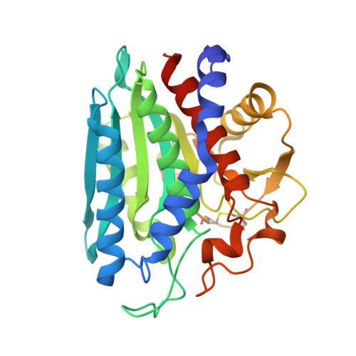
    3VH9

  • PubMed Abstract: 

    The selective inhibition of an aminopeptidase from Aeromonas proteolytica (AAP), a dinuclear Zn(2+) hydrolase, by 8-quinolinol (8-hydroxyquinoline, 8-HQ) derivatives is reported. We previously reported on the preparation of 8-HQ-pendant cyclens as Zn(2+) fluorophores (cyclen is 1,4,7,10-tetraazacyclododecane), in which the nitrogen and phenolate of the 8-HQ units (as well as the four nitrogens of cyclen) bind to Zn(2+) in a bidentate manner to form very stable Zn(2+) complexes at neutral pH (K (d) = 8-50 fM at pH 7.4). On the basis of this finding, it was hypothesized that 8-HQ derivatives have the potential to function as specific inhibitors of Zn(2+) enzymes, especially dinuclear Zn(2+) hydrolases. Assays of 8-HQ derivatives as inhibitors were performed against commercially available dinuclear Zn(2+) enzymes such as AAP and alkaline phosphatase. 8-HQ and the 5-substituted 8-HQ derivatives were found to be competitive inhibitors of AAP with inhibition constants of 0.16-29 μM at pH 8.0. The nitrogen at the 1-position and the hydroxide at the 8-position of 8-HQ were found to be essential for the inhibition of AAP. Fluorescence titrations of these drugs with AAP and an X-ray crystal structure analysis of an AAP-8-HQ complex (1.3-Å resolution) confirmed that 8-HQ binds to AAP in the "Pyr-out" mode, in which the hydroxide anion of 8-HQ bridges two Zn(2+) ions (Zn1 and Zn2) in the active site of AAP and the nitrogen atom of 8-HQ coordinates to Zn1 (Protein Data Bank code 3VH9).


  • Organizational Affiliation

    Faculty of Pharmaceutical Sciences, Tokyo University of Science, 2641 Yamazaki, Noda 278-8510, Japan.


Macromolecules
Find similar proteins by:  (by identity cutoff)  |  3D Structure
Entity ID: 1
MoleculeChains Sequence LengthOrganismDetailsImage
Bacterial leucyl aminopeptidase299Vibrio proteolyticusMutation(s): 0 
EC: 3.4.11.10
UniProt
Find proteins for Q01693 (Vibrio proteolyticus)
Explore Q01693 
Go to UniProtKB:  Q01693
Entity Groups  
Sequence Clusters30% Identity50% Identity70% Identity90% Identity95% Identity100% Identity
UniProt GroupQ01693
Sequence Annotations
Expand
  • Reference Sequence
Small Molecules
Ligands 6 Unique
IDChains Name / Formula / InChI Key2D Diagram3D Interactions
HQY
Query on HQY

Download Ideal Coordinates CCD File 
D [auth A]quinolin-8-ol
C9 H7 N O
MCJGNVYPOGVAJF-UHFFFAOYSA-N
GOL
Query on GOL

Download Ideal Coordinates CCD File 
Y [auth A]GLYCEROL
C3 H8 O3
PEDCQBHIVMGVHV-UHFFFAOYSA-N
ZN
Query on ZN

Download Ideal Coordinates CCD File 
B [auth A],
C [auth A]
ZINC ION
Zn
PTFCDOFLOPIGGS-UHFFFAOYSA-N
SCN
Query on SCN

Download Ideal Coordinates CCD File 
W [auth A],
X [auth A]
THIOCYANATE ION
C N S
ZMZDMBWJUHKJPS-UHFFFAOYSA-M
CL
Query on CL

Download Ideal Coordinates CCD File 
N [auth A]
O [auth A]
P [auth A]
Q [auth A]
R [auth A]
N [auth A],
O [auth A],
P [auth A],
Q [auth A],
R [auth A],
S [auth A],
T [auth A],
U [auth A],
V [auth A]
CHLORIDE ION
Cl
VEXZGXHMUGYJMC-UHFFFAOYSA-M
NA
Query on NA

Download Ideal Coordinates CCD File 
E [auth A]
F [auth A]
G [auth A]
H [auth A]
I [auth A]
E [auth A],
F [auth A],
G [auth A],
H [auth A],
I [auth A],
J [auth A],
K [auth A],
L [auth A],
M [auth A]
SODIUM ION
Na
FKNQFGJONOIPTF-UHFFFAOYSA-N
Binding Affinity Annotations 
IDSourceBinding Affinity
HQY PDBBind:  3VH9 Ki: 580 (nM) from 1 assay(s)
Binding MOAD:  3VH9 Ki: 580 (nM) from 1 assay(s)
Experimental Data & Validation

Experimental Data

  • Method: X-RAY DIFFRACTION
  • Resolution: 1.29 Å
  • R-Value Free: 0.153 
  • R-Value Work: 0.131 
  • R-Value Observed: 0.132 
  • Space Group: P 61 2 2
Unit Cell:
Length ( Å )Angle ( ˚ )
a = 108.463α = 90
b = 108.463β = 90
c = 90.737γ = 120
Software Package:
Software NamePurpose
HKL-2000data collection
PHASERphasing
PHENIXrefinement
REFMACrefinement
HKL-2000data reduction
HKL-2000data scaling

Structure Validation

View Full Validation Report


Entry History 

Deposition Data

Revision History  (Full details and data files)

  • Version 1.0: 2012-05-02
    Type: Initial release
  • Version 1.1: 2023-11-08
    Changes: Data collection, Database references, Derived calculations, Refinement description