3IHR

Crystal Structure of Uch37


Experimental Data Snapshot

  • Method: X-RAY DIFFRACTION
  • Resolution: 2.95 Å
  • R-Value Free: 0.242 
  • R-Value Work: 0.199 
  • R-Value Observed: 0.201 

wwPDB Validation   3D Report Full Report


This is version 1.8 of the entry. See complete history


Literature

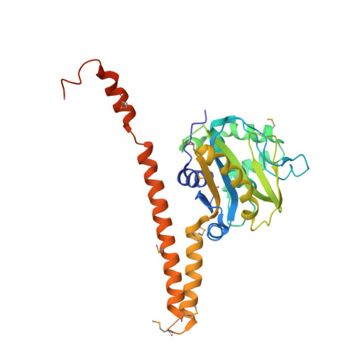
Structural characterization of human Uch37.

Burgie, S.E.Bingman, C.A.Soni, A.B.Phillips Jr., G.N.

(2012) Proteins 80: 649-654

  • DOI: https://doi.org/10.1002/prot.23147
  • Primary Citation of Related Structures:  
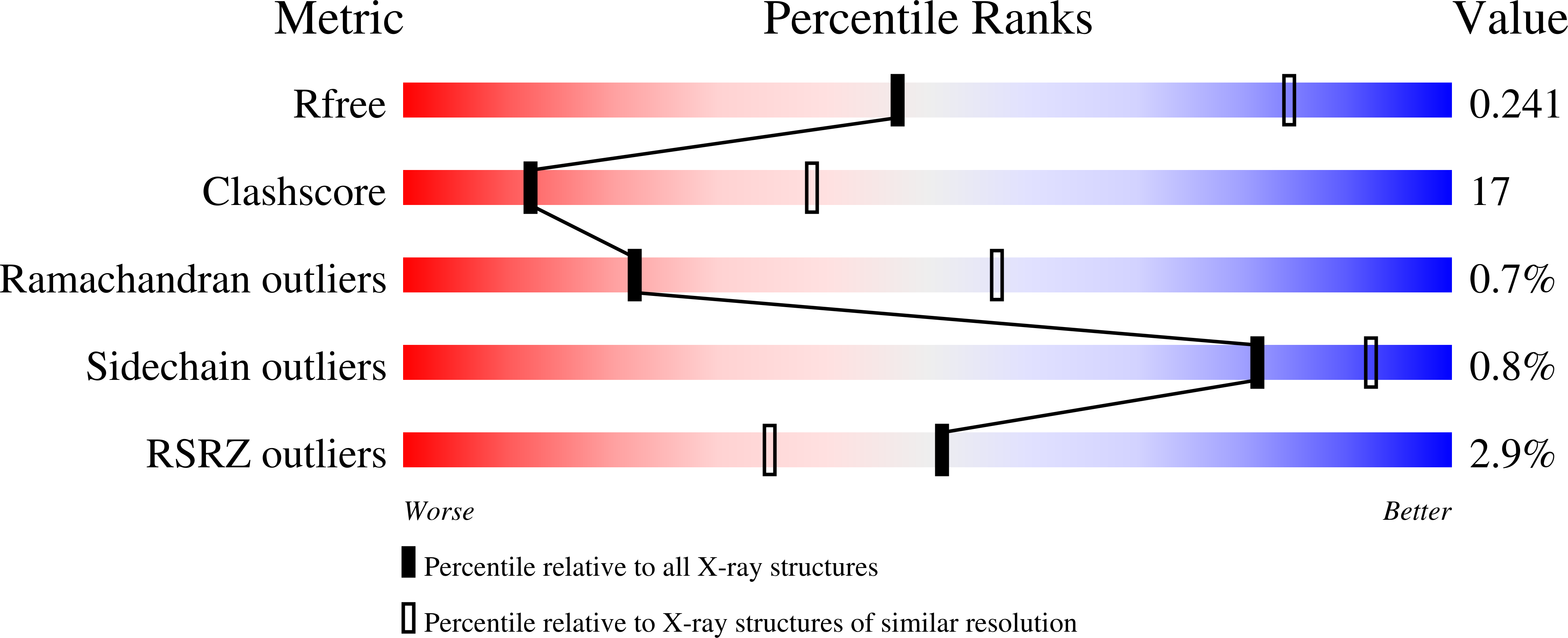
    3IHR

  • PubMed Abstract: 

    Uch37 is a de-ubiquitylating enzyme that is functionally linked with the 26S proteasome via Rpn13, and is essential for metazoan development. Here, we report the X-ray crystal structure of full-length human Uch37 at 2.95 Å resolution. Uch37's catalytic domain is similar to those of all UCH enzymes characterized to date. The C-terminal extension is elongated, predominantly helical and contains coiled coil interactions. Additionally, we provide an initial characterization of Uch37's oligomeric state and identify a systematic error in previous analyses of Uch37 activity. Taken together, these data provide a strong foundation for further analysis of Uch37's several functions.


  • Organizational Affiliation

    Department of Biochemistry, Center for Eukaryotic Structural Genomics, University of Wisconsin-Madison, Madison Wisconsin 53706-1544.


Macromolecules
Find similar proteins by:  (by identity cutoff)  |  3D Structure
Entity ID: 1
MoleculeChains Sequence LengthOrganismDetailsImage
Ubiquitin carboxyl-terminal hydrolase isozyme L5328Homo sapiensMutation(s): 1 
Gene Names: AD-019CGI-70UCH37UCHL5
EC: 3.4.19.12
UniProt & NIH Common Fund Data Resources
Find proteins for Q9Y5K5 (Homo sapiens)
Explore Q9Y5K5 
Go to UniProtKB:  Q9Y5K5
PHAROS:  Q9Y5K5
GTEx:  ENSG00000116750 
Entity Groups  
Sequence Clusters30% Identity50% Identity70% Identity90% Identity95% Identity100% Identity
UniProt GroupQ9Y5K5
Sequence Annotations
Expand
  • Reference Sequence
Small Molecules
Modified Residues  1 Unique
IDChains TypeFormula2D DiagramParent
MSE
Query on MSE
A
L-PEPTIDE LINKINGC5 H11 N O2 SeMET
Experimental Data & Validation

Experimental Data

  • Method: X-RAY DIFFRACTION
  • Resolution: 2.95 Å
  • R-Value Free: 0.242 
  • R-Value Work: 0.199 
  • R-Value Observed: 0.201 
  • Space Group: I 2 2 2
Unit Cell:
Length ( Å )Angle ( ˚ )
a = 90.092α = 90
b = 98.736β = 90
c = 154.061γ = 90
Software Package:
Software NamePurpose
DENZOdata reduction
SCALEPACKdata scaling
SHARPphasing
DMphasing
PHENIXrefinement
PDB_EXTRACTdata extraction
HKL-2000data collection
HKL-2000data reduction
HKL-2000data scaling

Structure Validation

View Full Validation Report


Entry History 

Deposition Data

Revision History  (Full details and data files)

  • Version 1.0: 2009-08-11
    Type: Initial release
  • Version 1.1: 2011-07-13
    Changes: Version format compliance
  • Version 1.2: 2012-10-10
    Changes: Database references
  • Version 1.3: 2013-01-09
    Changes: Database references
  • Version 1.4: 2013-01-23
    Changes: Database references
  • Version 1.5: 2013-06-19
    Changes: Database references
  • Version 1.6: 2017-11-01
    Changes: Refinement description
  • Version 1.7: 2021-10-13
    Changes: Database references, Derived calculations
  • Version 1.8: 2023-07-26
    Changes: Database references