3ZYR

Structure of the lectin from Platypodium elegans in complex with heptasaccharide


Experimental Data Snapshot

  • Method: X-RAY DIFFRACTION
  • Resolution: 1.65 Å
  • R-Value Free: 0.188 
  • R-Value Work: 0.156 
  • R-Value Observed: 0.157 

wwPDB Validation   3D Report Full Report


This is version 2.0 of the entry. See complete history


Literature

A Lectin from Platypodium Elegans with Unusual Specificity and Affinity for Asymmetric Complex N-Glycans.

Benevides, R.G.Ganne, G.Simoes, R.D.C.Schubert, V.Niemietz, M.Unverzagt, C.Chazalet, V.Breton, C.Varrot, A.Cavada, B.S.Imberty, A.

(2012) J Biol Chem 287: 26352

  • DOI: https://doi.org/10.1074/jbc.M112.375816
  • Primary Citation of Related Structures:  
    3ZVX, 3ZYR

  • PubMed Abstract: 

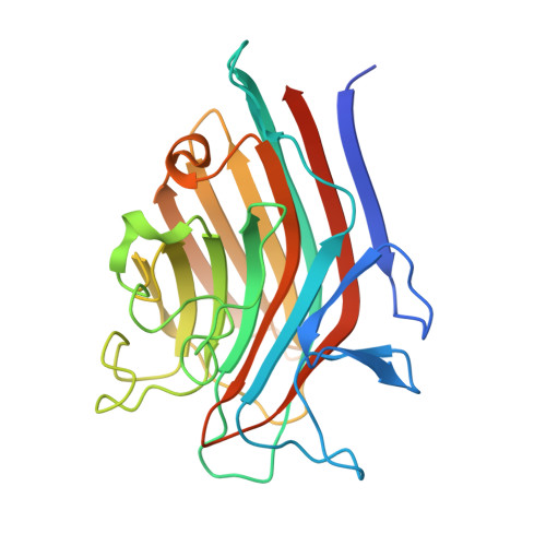
    Lectin activity with specificity for mannose and glucose has been detected in the seed of Platypodium elegans, a legume plant from the Dalbergieae tribe. The gene of Platypodium elegans lectin A has been cloned, and the resulting 261-amino acid protein belongs to the legume lectin family with similarity with Pterocarpus angolensis agglutinin from the same tribe. The recombinant lectin has been expressed in Escherichia coli and refolded from inclusion bodies. Analysis of specificity by glycan array evidenced a very unusual preference for complex type N-glycans with asymmetrical branches. A short branch consisting of one mannose residue is preferred on the 6-arm of the N-glycan, whereas extensions by GlcNAc, Gal, and NeuAc are favorable on the 3-arm. Affinities have been obtained by microcalorimetry using symmetrical and asymmetrical Asn-linked heptasaccharides prepared by the semi-synthetic method. Strong affinity with K(d) of 4.5 μm was obtained for both ligands. Crystal structures of Platypodium elegans lectin A complexed with branched trimannose and symmetrical complex-type Asn-linked heptasaccharide have been solved at 2.1 and 1.65 Å resolution, respectively. The lectin adopts the canonical dimeric organization of legume lectins. The trimannose bridges the binding sites of two neighboring dimers, resulting in the formation of infinite chains in the crystal. The Asn-linked heptasaccharide binds with the 6-arm in the primary binding site with extensive additional contacts on both arms. The GlcNAc on the 6-arm is bound in a constrained conformation that may rationalize the higher affinity observed on the glycan array for N-glycans with only a mannose on the 6-arm.


  • Organizational Affiliation

    Centre de Recherche sur les Macromolécules Végétales-CNRS (affiliated with Université Joseph Fourier and Institut de Chimie Moléculaire de Grenoble), 38041 Grenoble, France.


Macromolecules
Find similar proteins by:  (by identity cutoff)  |  3D Structure
Entity ID: 1
MoleculeChains Sequence LengthOrganismDetailsImage
LECTIN
A, B
261Platypodium elegansMutation(s): 0 
UniProt
Find proteins for G1EUI6 (Platypodium elegans)
Explore G1EUI6 
Go to UniProtKB:  G1EUI6
Entity Groups  
Sequence Clusters30% Identity50% Identity70% Identity90% Identity95% Identity100% Identity
UniProt GroupG1EUI6
Sequence Annotations
Expand
  • Reference Sequence
Oligosaccharides

Help

Entity ID: 2
MoleculeChains Length2D Diagram Glycosylation3D Interactions
2-acetamido-2-deoxy-beta-D-glucopyranose-(1-2)-alpha-D-mannopyranose-(1-3)-[2-acetamido-2-deoxy-beta-D-glucopyranose-(1-2)-alpha-D-mannopyranose-(1-6)]beta-D-mannopyranose-(1-4)-2-acetamido-2-deoxy-beta-D-glucopyranose-(1-4)-2-acetamido-2-deoxy-beta-D-glucopyranose
C, D
7N-Glycosylation
Glycosylation Resources
GlyTouCan:  G39213VZ
GlyCosmos:  G39213VZ
GlyGen:  G39213VZ
Small Molecules
Ligands 5 Unique
IDChains Name / Formula / InChI Key2D Diagram3D Interactions
ASN
Query on ASN

Download Ideal Coordinates CCD File 
E [auth A]ASPARAGINE
C4 H8 N2 O3
DCXYFEDJOCDNAF-REOHCLBHSA-N
GOL
Query on GOL

Download Ideal Coordinates CCD File 
H [auth A],
J [auth A],
K [auth A],
N [auth B]
GLYCEROL
C3 H8 O3
PEDCQBHIVMGVHV-UHFFFAOYSA-N
URE
Query on URE

Download Ideal Coordinates CCD File 
I [auth A],
O [auth B]
UREA
C H4 N2 O
XSQUKJJJFZCRTK-UHFFFAOYSA-N
MN
Query on MN

Download Ideal Coordinates CCD File 
F [auth A],
L [auth B]
MANGANESE (II) ION
Mn
WAEMQWOKJMHJLA-UHFFFAOYSA-N
CA
Query on CA

Download Ideal Coordinates CCD File 
G [auth A],
M [auth B]
CALCIUM ION
Ca
BHPQYMZQTOCNFJ-UHFFFAOYSA-N
Experimental Data & Validation

Experimental Data

  • Method: X-RAY DIFFRACTION
  • Resolution: 1.65 Å
  • R-Value Free: 0.188 
  • R-Value Work: 0.156 
  • R-Value Observed: 0.157 
  • Space Group: P 21 21 21
Unit Cell:
Length ( Å )Angle ( ˚ )
a = 51.4α = 90
b = 76.98β = 90
c = 125.59γ = 90
Software Package:
Software NamePurpose
REFMACrefinement
MOSFLMdata reduction
SCALAdata scaling
PHASERphasing

Structure Validation

View Full Validation Report


Entry History 

Deposition Data

Revision History  (Full details and data files)

  • Version 1.0: 2012-01-11
    Type: Initial release
  • Version 1.1: 2012-01-25
    Changes: Other
  • Version 1.2: 2012-07-25
    Changes: Database references, Structure summary
  • Version 1.3: 2012-08-08
    Changes: Database references
  • Version 2.0: 2020-07-29
    Type: Remediation
    Reason: Carbohydrate remediation
    Changes: Advisory, Atomic model, Data collection, Derived calculations, Other, Structure summary