3WB8

Crystal Structure of MyoVa-GTD


Experimental Data Snapshot

  • Method: X-RAY DIFFRACTION
  • Resolution: 2.50 Å
  • R-Value Free: 0.225 
  • R-Value Work: 0.173 
  • R-Value Observed: 0.176 

wwPDB Validation   3D Report Full Report


This is version 1.2 of the entry. See complete history


Literature

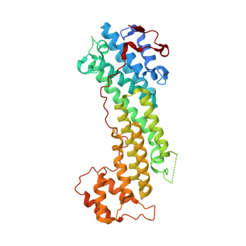
Structural basis of cargo recognitions for class V myosins

Wei, Z.Liu, X.Yu, C.Zhang, M.

(2013) Proc Natl Acad Sci U S A 110: 11314-11319

  • DOI: https://doi.org/10.1073/pnas.1306768110
  • Primary Citation of Related Structures:  
    3WB8, 4KP3

  • PubMed Abstract: 

    Class V myosins (MyoV), the most studied unconventional myosins, recognize numerous cargos mainly via the motor's globular tail domain (GTD). Little is known regarding how MyoV-GTD recognizes such a diverse array of cargos specifically. Here, we solved the crystal structures of MyoVa-GTD in its apo-form and in complex with two distinct cargos, melanophilin and Rab interacting lysosomal protein-like 2. The apo-MyoVa-GTD structure indicates that most mutations found in patients with Griscelli syndrome, microvillus inclusion disease, or cancers or in "dilute" rodents likely impair the folding of GTD. The MyoVa-GTD/cargo complex structure reveals two distinct cargo-binding surfaces, one primarily via charge-charge interaction and the other mainly via hydrophobic interactions. Structural and biochemical analysis reveal the specific cargo-binding specificities of various isoforms of mammalian MyoV as well as very different cargo recognition mechanisms of MyoV between yeast and higher eukaryotes. The MyoVa-GTD structures resolved here provide a framework for future functional studies of vertebrate class V myosins.


  • Organizational Affiliation

    Division of Life Science, State Key Laboratory of Molecular Neuroscience, Hong Kong University of Science and Technology, Kowloon, Hong Kong, China.


Macromolecules
Find similar proteins by:  (by identity cutoff)  |  3D Structure
Entity ID: 1
MoleculeChains Sequence LengthOrganismDetailsImage
Unconventional myosin-Va
A, B, C, D, E
A, B, C, D, E, F, G, H
404Mus musculusMutation(s): 0 
Gene Names: Myo5a
UniProt & NIH Common Fund Data Resources
Find proteins for Q99104 (Mus musculus)
Explore Q99104 
Go to UniProtKB:  Q99104
IMPC:  MGI:105976
Entity Groups  
Sequence Clusters30% Identity50% Identity70% Identity90% Identity95% Identity100% Identity
UniProt GroupQ99104
Sequence Annotations
Expand
  • Reference Sequence
Small Molecules
Ligands 1 Unique
IDChains Name / Formula / InChI Key2D Diagram3D Interactions
EDO
Query on EDO

Download Ideal Coordinates CCD File 
AA [auth G]
BA [auth G]
CA [auth H]
DA [auth H]
I [auth A]
AA [auth G],
BA [auth G],
CA [auth H],
DA [auth H],
I [auth A],
J [auth A],
K [auth A],
L [auth A],
M [auth B],
N [auth B],
O [auth C],
P [auth C],
Q [auth C],
R [auth D],
S [auth D],
T [auth D],
U [auth E],
V [auth E],
W [auth F],
X [auth F],
Y [auth F],
Z [auth G]
1,2-ETHANEDIOL
C2 H6 O2
LYCAIKOWRPUZTN-UHFFFAOYSA-N
Experimental Data & Validation

Experimental Data

  • Method: X-RAY DIFFRACTION
  • Resolution: 2.50 Å
  • R-Value Free: 0.225 
  • R-Value Work: 0.173 
  • R-Value Observed: 0.176 
  • Space Group: P 1 21 1
Unit Cell:
Length ( Å )Angle ( ˚ )
a = 68.984α = 90
b = 165.355β = 90.06
c = 173.02γ = 90
Software Package:
Software NamePurpose
PHENIXrefinement
PDB_EXTRACTdata extraction
Blu-Icedata collection
HKL-2000data reduction
HKL-2000data scaling
AutoSolphasing

Structure Validation

View Full Validation Report


Entry History 

Deposition Data

Revision History  (Full details and data files)

  • Version 1.0: 2013-07-10
    Type: Initial release
  • Version 1.1: 2013-08-14
    Changes: Database references
  • Version 1.2: 2024-03-20
    Changes: Data collection, Database references, Derived calculations