Crystal Structure of Bacillus subtilis Signal Peptide Peptidase A.
Nam, S.E., Kim, A.C., Paetzel, M.(2012) J Mol Biol 419: 347-358
- PubMed: 22472423
- DOI: https://doi.org/10.1016/j.jmb.2012.03.020
- Primary Citation of Related Structures:
3RST - PubMed Abstract:
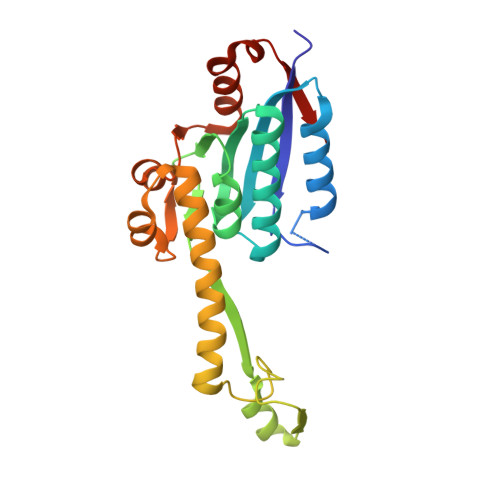
Signal peptide peptidase A (SppA) is a membrane-bound self-compartmentalized serine protease that functions to cleave the remnant signal peptides left behind after protein secretion and cleavage by signal peptidases. SppA is found in plants, archaea and bacteria. Here, we report the first crystal structure of a Gram-positive bacterial SppA. The 2.4-Å-resolution structure of Bacillus subtilis SppA (SppA(BS)) catalytic domain reveals eight SppA(BS) molecules in the asymmetric unit, forming a dome-shaped octameric complex. The octameric state of SppA(BS) is supported by analytical size-exclusion chromatography and multi-angle light scattering analysis. Our sequence analysis, mutagenesis and activity assays are consistent with Ser147 serving as the nucleophile and Lys199 serving as the general base; however, they are located in different region of the protein, more than 29 Å apart. Only upon assembling the octamer do the serine and lysine come within close proximity, with neighboring protomers each providing one-half of the catalytic dyad, thus producing eight separate active sites within the complex, twice the number seen within Escherichia coli SppA (SppA(EC)). The SppA(BS) S1 substrate specificity pocket is deep, narrow and hydrophobic, but with a polar bottom. The S3 pocket, which is constructed from two neighboring proteins, is shallower, wider and more polar than the S1 pocket. A comparison of these pockets to those seen in SppA(EC) reveals a significant difference in the size and shape of the S1 pocket, which we show is reflected in the repertoire of peptides the enzymes are capable of cleaving.
Organizational Affiliation:
Department of Molecular Biology and Biochemistry, Simon Fraser University, South Science Building 8888 University Drive, Burnaby, British Columbia, Canada V5A 1S6.