3R4D

Crystal structure of mouse coronavirus receptor-binding domain complexed with its murine receptor


Experimental Data Snapshot

  • Method: X-RAY DIFFRACTION
  • Resolution: 3.10 Å
  • R-Value Free: 0.308 
  • R-Value Work: 0.248 

wwPDB Validation   3D Report Full Report


Ligand Structure Quality Assessment 


This is version 2.0 of the entry. See complete history


Literature

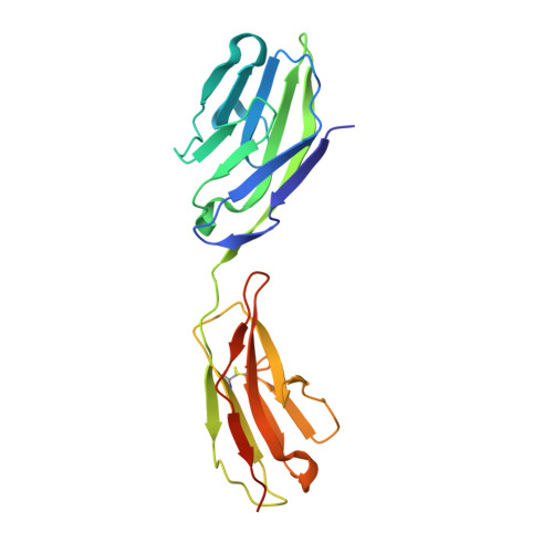
Crystal structure of mouse coronavirus receptor-binding domain complexed with its murine receptor.

Peng, G.Sun, D.Rajashankar, K.R.Qian, Z.Holmes, K.V.Li, F.

(2011) Proc Natl Acad Sci U S A 108: 10696-10701

  • DOI: https://doi.org/10.1073/pnas.1104306108
  • Primary Citation of Related Structures:  
    3R4D

  • PubMed Abstract: 

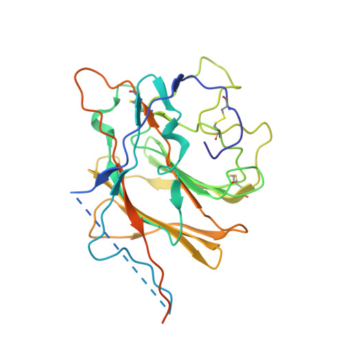
    Coronaviruses have evolved diverse mechanisms to recognize different receptors for their cross-species transmission and host-range expansion. Mouse hepatitis coronavirus (MHV) uses the N-terminal domain (NTD) of its spike protein as its receptor-binding domain. Here we present the crystal structure of MHV NTD complexed with its receptor murine carcinoembryonic antigen-related cell adhesion molecule 1a (mCEACAM1a). Unexpectedly, MHV NTD contains a core structure that has the same β-sandwich fold as human galectins (S-lectins) and additional structural motifs that bind to the N-terminal Ig-like domain of mCEACAM1a. Despite its galectin fold, MHV NTD does not bind sugars, but instead binds mCEACAM1a through exclusive protein-protein interactions. Critical contacts at the interface have been confirmed by mutagenesis, providing a structural basis for viral and host specificities of coronavirus/CEACAM1 interactions. Sugar-binding assays reveal that galectin-like NTDs of some coronaviruses such as human coronavirus OC43 and bovine coronavirus bind sugars. Structural analysis and mutagenesis localize the sugar-binding site in coronavirus NTDs to be above the β-sandwich core. We propose that coronavirus NTDs originated from a host galectin and retained sugar-binding functions in some contemporary coronaviruses, but evolved new structural features in MHV for mCEACAM1a binding.


  • Organizational Affiliation

    Department of Pharmacology, University of Minnesota Medical School, Minneapolis, MN 55455, USA.


Macromolecules
Find similar proteins by:  (by identity cutoff)  |  3D Structure
Entity ID: 1
MoleculeChains Sequence LengthOrganismDetailsImage
CEA-related cell adhesion molecule 1, isoform 1/2S
A, C
208Mus musculusMutation(s): 0 
Gene Names: Ceacam1CEACAM1a-2S
UniProt
Find proteins for P31809 (Mus musculus)
Explore P31809 
Go to UniProtKB:  P31809
Entity Groups  
Sequence Clusters30% Identity50% Identity70% Identity90% Identity95% Identity100% Identity
UniProt GroupP31809
Sequence Annotations
Expand
  • Reference Sequence
Find similar proteins by:  (by identity cutoff)  |  3D Structure
Entity ID: 2
MoleculeChains Sequence LengthOrganismDetailsImage
Spike glycoprotein
B, D
288Murine coronavirusMutation(s): 0 
UniProt
Find proteins for Q9J3E7 (Murine hepatitis virus)
Explore Q9J3E7 
Go to UniProtKB:  Q9J3E7
Entity Groups  
Sequence Clusters30% Identity50% Identity70% Identity90% Identity95% Identity100% Identity
UniProt GroupQ9J3E7
Sequence Annotations
Expand
  • Reference Sequence
Oligosaccharides

Help

Entity ID: 3
MoleculeChains Length2D Diagram Glycosylation3D Interactions
2-acetamido-2-deoxy-beta-D-glucopyranose-(1-4)-2-acetamido-2-deoxy-beta-D-glucopyranose
E, F
2N-Glycosylation
Glycosylation Resources
GlyTouCan:  G42666HT
GlyCosmos:  G42666HT
GlyGen:  G42666HT
Experimental Data & Validation

Experimental Data

  • Method: X-RAY DIFFRACTION
  • Resolution: 3.10 Å
  • R-Value Free: 0.308 
  • R-Value Work: 0.248 
  • Space Group: P 61 2 2
Unit Cell:
Length ( Å )Angle ( ˚ )
a = 76.38α = 90
b = 76.38β = 90
c = 942.09γ = 120
Software Package:
Software NamePurpose
HKL-3000data collection
SOLVEphasing
REFMACrefinement
HKL-3000data reduction
HKL-3000data scaling

Structure Validation

View Full Validation Report


Ligand Structure Quality Assessment 


Entry History 

Deposition Data

Revision History  (Full details and data files)

  • Version 1.0: 2011-06-22
    Type: Initial release
  • Version 1.1: 2011-07-13
    Changes: Version format compliance
  • Version 1.2: 2014-10-29
    Changes: Structure summary
  • Version 1.3: 2020-04-15
    Changes: Data collection, Database references, Derived calculations, Source and taxonomy, Structure summary
  • Version 2.0: 2020-07-29
    Type: Remediation
    Reason: Carbohydrate remediation
    Changes: Atomic model, Data collection, Derived calculations, Structure summary