3PMH

Mechanism of Sulfotyrosine-Mediated Glycoprotein Ib Interaction with Two Distinct alpha-Thrombin Sites


Experimental Data Snapshot

  • Method: X-RAY DIFFRACTION
  • Resolution: 3.20 Å
  • R-Value Free: 0.263 
  • R-Value Work: 0.220 
  • R-Value Observed: 0.220 

wwPDB Validation   3D Report Full Report


Ligand Structure Quality Assessment 


This is version 2.2 of the entry. See complete history


Literature

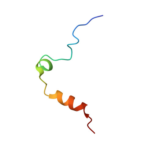
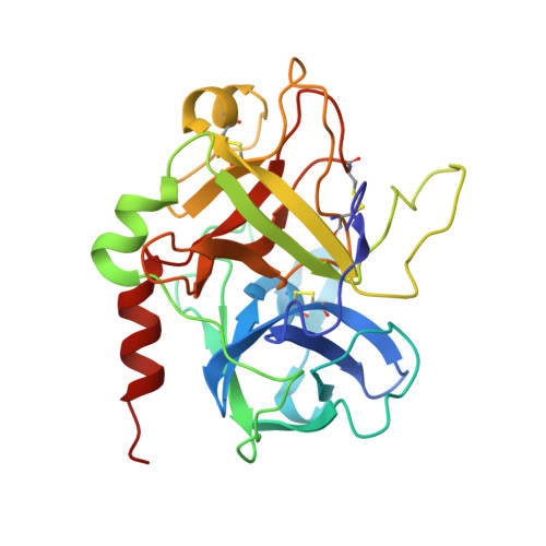
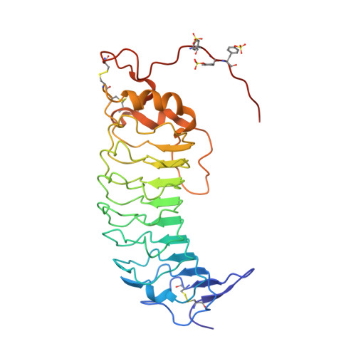
Binding of alpha-thrombin to surface-anchored platelet glycoprotein Ib(alpha) sulfotyrosines through a two-site mechanism involving exosite I.

Zarpellon, A.Celikel, R.Roberts, J.R.McClintock, R.A.Mendolicchio, G.L.Moore, K.L.Jing, H.Varughese, K.I.Ruggeri, Z.M.

(2011) Proc Natl Acad Sci U S A 108: 8628-8633

  • DOI: https://doi.org/10.1073/pnas.1017042108
  • Primary Citation of Related Structures:  
    3PMH

  • PubMed Abstract: 

    The involvement of exosite I in α-thrombin (FIIa) binding to platelet glycoprotein Ibα (GPIbα), which could influence interactions with other substrates, remains undefined. To address the problem, we generated the GPIbα amino terminal domain (GPIbα-N) fully sulfated on three tyrosine residues and solved the structure of its complex with FIIa. We found that sulfotyrosine (Tys) 278 enhances the interaction mainly by establishing contacts with exosite I. We then evaluated how substituting tyrosine with phenylalanine, which cannot be sulfated, affects FIIa binding to soluble or surface-immobilized GPIbα-N. Mutating Tyr(276), which mostly contacts exosite II residues, markedly reduced FIIa interaction with both soluble and immobilized GPIbα-N; mutating Tyr(278) or Tyr(279), which mostly contact exosite I residues, reduced FIIa complexing in solution by 0-20% but affinity for immobilized GPIbα-N 2 to 6-fold, respectively. Moreover, three exosite I ligands--aptamer HD1, hirugen, and lepirudin--did not interfere with soluble FIIa complexing to GPIbα-N, excluding that their binding caused allosteric effects influencing the interaction; nonetheless, all impaired FIIa binding to immobilized GPIbα-N and platelet GPIb nearly as much as aptamer HD22 and heparin, both exosite II ligands. Bound HD1 and hirugen alter Trp(148) orientation in a loop near exosite I preventing contacts with the sulfate oxygen atoms of Tys(279). These results support a mechanism in which binding occurs when the two exosites of one FIIa molecule independently interact with two immobilized GPIbα molecules. Through exosite engagement, GPIbα may influence FIIa-dependent processes relevant to hemostasis and thrombosis.


  • Organizational Affiliation

    Department of Molecular and Experimental Medicine, Roon Research Center for Arteriosclerosis and Thrombosis, The Scripps Research Institute, La Jolla, CA 92037, USA.


Macromolecules
Find similar proteins by:  (by identity cutoff)  |  3D Structure
Entity ID: 1
MoleculeChains Sequence LengthOrganismDetailsImage
THROMBIN ALPHA-CHAIN36Homo sapiensMutation(s): 0 
EC: 3.4.21.5
UniProt & NIH Common Fund Data Resources
Find proteins for P00734 (Homo sapiens)
Explore P00734 
Go to UniProtKB:  P00734
PHAROS:  P00734
GTEx:  ENSG00000180210 
Entity Groups  
Sequence Clusters30% Identity50% Identity70% Identity90% Identity95% Identity100% Identity
UniProt GroupP00734
Sequence Annotations
Expand
  • Reference Sequence
Find similar proteins by:  (by identity cutoff)  |  3D Structure
Entity ID: 2
MoleculeChains Sequence LengthOrganismDetailsImage
THROMBIN BETA-CHAIN259Homo sapiensMutation(s): 0 
EC: 3.4.21.5
UniProt & NIH Common Fund Data Resources
Find proteins for P00734 (Homo sapiens)
Explore P00734 
Go to UniProtKB:  P00734
PHAROS:  P00734
GTEx:  ENSG00000180210 
Entity Groups  
Sequence Clusters30% Identity50% Identity70% Identity90% Identity95% Identity100% Identity
UniProt GroupP00734
Sequence Annotations
Expand
  • Reference Sequence
Find similar proteins by:  (by identity cutoff)  |  3D Structure
Entity ID: 3
MoleculeChains Sequence LengthOrganismDetailsImage
Platelet glycoprotein Ib alpha chainC [auth G]290Homo sapiensMutation(s): 1 
Gene Names: GP1BAplatelet glycoprotein Ib alpha
UniProt & NIH Common Fund Data Resources
Find proteins for P07359 (Homo sapiens)
Explore P07359 
Go to UniProtKB:  P07359
PHAROS:  P07359
GTEx:  ENSG00000185245 
Entity Groups  
Sequence Clusters30% Identity50% Identity70% Identity90% Identity95% Identity100% Identity
UniProt GroupP07359
Sequence Annotations
Expand
  • Reference Sequence
Oligosaccharides

Help

Entity ID: 4
MoleculeChains Length2D Diagram Glycosylation3D Interactions
alpha-D-mannopyranose-(1-3)-[alpha-D-mannopyranose-(1-6)]alpha-D-mannopyranose-(1-4)-2-acetamido-2-deoxy-beta-D-glucopyranose-(1-4)-2-acetamido-2-deoxy-beta-D-glucopyranoseD [auth C]5N-Glycosylation
Glycosylation Resources
GlyTouCan:  G21381MC
GlyCosmos:  G21381MC
GlyGen:  G21381MC
Small Molecules
Ligands 2 Unique
IDChains Name / Formula / InChI Key2D Diagram3D Interactions
0G7
Query on 0G7

Download Ideal Coordinates CCD File 
E [auth B]D-phenylalanyl-N-[(3S)-6-carbamimidamido-1-chloro-2-oxohexan-3-yl]-L-prolinamide
C21 H31 Cl N6 O3
KWPACVJPAFGBEQ-IKGGRYGDSA-N
NAG
Query on NAG

Download Ideal Coordinates CCD File 
F [auth G]2-acetamido-2-deoxy-beta-D-glucopyranose
C8 H15 N O6
OVRNDRQMDRJTHS-FMDGEEDCSA-N
Modified Residues  1 Unique
IDChains TypeFormula2D DiagramParent
TYS
Query on TYS
C [auth G]L-PEPTIDE LINKINGC9 H11 N O6 STYR
Binding Affinity Annotations 
IDSourceBinding Affinity
0G7 BindingDB:  3PMH IC50: 1.5 (nM) from 1 assay(s)
Biologically Interesting Molecules (External Reference) 1 Unique
Experimental Data & Validation

Experimental Data

  • Method: X-RAY DIFFRACTION
  • Resolution: 3.20 Å
  • R-Value Free: 0.263 
  • R-Value Work: 0.220 
  • R-Value Observed: 0.220 
  • Space Group: P 43 21 2
Unit Cell:
Length ( Å )Angle ( ˚ )
a = 67.657α = 90
b = 67.657β = 90
c = 329.73γ = 90
Software Package:
Software NamePurpose
ADSCdata collection
CNSrefinement
HKL-2000data reduction
SCALEPACKdata scaling
CNSphasing

Structure Validation

View Full Validation Report


Ligand Structure Quality Assessment 


Entry History 

Deposition Data

Revision History  (Full details and data files)

  • Version 1.0: 2011-06-01
    Type: Initial release
  • Version 1.1: 2011-07-13
    Changes: Version format compliance
  • Version 1.2: 2011-08-10
    Changes: Database references
  • Version 1.3: 2012-12-12
    Changes: Other
  • Version 2.0: 2020-07-29
    Type: Remediation
    Reason: Carbohydrate remediation
    Changes: Atomic model, Data collection, Database references, Derived calculations, Structure summary
  • Version 2.1: 2023-09-06
    Changes: Data collection, Database references, Refinement description, Structure summary
  • Version 2.2: 2023-12-06
    Changes: Data collection