3M66

Crystal structure of human Mitochondrial Transcription Termination Factor 3


Experimental Data Snapshot

  • Method: X-RAY DIFFRACTION
  • Resolution: 1.60 Å
  • R-Value Free: 0.199 
  • R-Value Work: 0.173 
  • R-Value Observed: 0.175 

wwPDB Validation   3D Report Full Report


This is version 1.2 of the entry. See complete history


Literature

Structure of mitochondrial transcription termination factor 3 reveals a novel nucleic acid-binding domain.

Spahr, H.Samuelsson, T.Hallberg, B.M.Gustafsson, C.M.

(2010) Biochem Biophys Res Commun 397: 386-390

  • DOI: https://doi.org/10.1016/j.bbrc.2010.04.130
  • Primary Citation of Related Structures:  
    3M66

  • PubMed Abstract: 

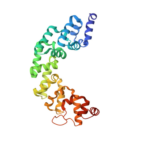
    In mammalian cells, a family of mitochondrial transcription termination factors (MTERFs) regulates mitochondrial gene expression. MTERF family members share a approximately 270 residues long MTERF-domain required for DNA binding and transcription regulation. However, the structure of this widely conserved domain is unknown. Here, we show that the MTERF-domain of human MTERF3 forms a half-doughnut-shaped right-handed superhelix. The superhelix is built from alpha-helical tandem repeats that display a novel triangular three-helix motif. This repeat motif, which we denote the MTERF-motif, is a conserved structural element present in proteins from metazoans, plants, and protozoans. Furthermore, a narrow, strongly positively charged nucleic acid-binding path is found in the middle of the concave side of the half-doughnut. This arrangement suggests a half clamp nucleic acid-binding mode for MTERF-domains.


  • Organizational Affiliation

    Dept. of Laboratory Medicine, Division of Metabolic Diseases, Karolinska Institutet, Stockholm, Sweden.


Macromolecules
Find similar proteins by:  (by identity cutoff)  |  3D Structure
Entity ID: 1
MoleculeChains Sequence LengthOrganismDetailsImage
mTERF domain-containing protein 1, mitochondrial270Homo sapiensMutation(s): 0 
Gene Names: CGI-12MTERFD1
UniProt & NIH Common Fund Data Resources
Find proteins for Q96E29 (Homo sapiens)
Explore Q96E29 
Go to UniProtKB:  Q96E29
PHAROS:  Q96E29
GTEx:  ENSG00000156469 
Entity Groups  
Sequence Clusters30% Identity50% Identity70% Identity90% Identity95% Identity100% Identity
UniProt GroupQ96E29
Sequence Annotations
Expand
  • Reference Sequence
Experimental Data & Validation

Experimental Data

  • Method: X-RAY DIFFRACTION
  • Resolution: 1.60 Å
  • R-Value Free: 0.199 
  • R-Value Work: 0.173 
  • R-Value Observed: 0.175 
  • Space Group: C 1 2 1
Unit Cell:
Length ( Å )Angle ( ˚ )
a = 139.23α = 90
b = 32.24β = 106.59
c = 70.22γ = 90
Software Package:
Software NamePurpose
PHENIXmodel building
PHENIXrefinement
XDSdata reduction
XSCALEdata scaling
PHENIXphasing

Structure Validation

View Full Validation Report


Entry History 

Deposition Data

Revision History  (Full details and data files)

  • Version 1.0: 2010-05-05
    Type: Initial release
  • Version 1.1: 2011-07-13
    Changes: Version format compliance
  • Version 1.2: 2024-02-21
    Changes: Data collection, Database references