3ITK

Crystal structure of human UDP-glucose dehydrogenase Thr131Ala, apo form.


Experimental Data Snapshot

  • Method: X-RAY DIFFRACTION
  • Resolution: 2.40 Å
  • R-Value Free: 0.241 
  • R-Value Work: 0.198 
  • R-Value Observed: 0.199 

wwPDB Validation   3D Report Full Report


This is version 1.4 of the entry. See complete history


Literature

Structure and mechanism of human UDP-glucose 6-dehydrogenase.

Egger, S.Chaikuad, A.Kavanagh, K.L.Oppermann, U.Nidetzky, B.

(2011) J Biol Chem 286: 23877-23887

  • DOI: https://doi.org/10.1074/jbc.M111.234682
  • Primary Citation of Related Structures:  
    2Q3E, 2QG4, 3ITK

  • PubMed Abstract: 

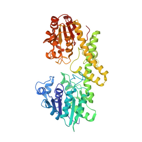
    Elevated production of the matrix glycosaminoglycan hyaluronan is strongly implicated in epithelial tumor progression. Inhibition of synthesis of the hyaluronan precursor UDP-glucuronic acid (UDP-GlcUA) therefore presents an emerging target for cancer therapy. Human UDP-glucose 6-dehydrogenase (hUGDH) catalyzes, in two NAD(+)-dependent steps without release of intermediate aldehyde, the biosynthetic oxidation of UDP-glucose (UDP-Glc) to UDP-GlcUA. Here, we present a structural characterization of the hUGDH reaction coordinate using crystal structures of the apoenzyme and ternary complexes of the enzyme bound with UDP-Glc/NADH and UDP-GlcUA/NAD(+). The quaternary structure of hUGDH is a disc-shaped trimer of homodimers whose subunits consist of two discrete α/β domains with the active site located in the interdomain cleft. Ternary complex formation is accompanied by rigid-body and restrained movement of the N-terminal NAD(+) binding domain, sequestering substrate and coenzyme in their reactive positions through interdomain closure. By alternating between conformations in and out of the active site during domain motion, Tyr(14), Glu(161), and Glu(165) participate in control of coenzyme binding and release during 2-fold oxidation. The proposed mechanism of hUGDH involves formation and breakdown of thiohemiacetal and thioester intermediates whereby Cys(276) functions as the catalytic nucleophile. Stopped-flow kinetic data capture the essential deprotonation of Cys(276) in the course of the first oxidation step, allowing the thiolate side chain to act as a trap of the incipient aldehyde. Because thiohemiacetal intermediate accumulates at steady state under physiological reaction conditions, hUGDH inhibition might best explore ligand binding to the NAD(+) binding domain.


  • Organizational Affiliation

    Institute of Biotechnology and Biochemical Engineering, Graz University of Technology, Petersgasse 12/1, A-8010 Graz, Austria.


Macromolecules
Find similar proteins by:  (by identity cutoff)  |  3D Structure
Entity ID: 1
MoleculeChains Sequence LengthOrganismDetailsImage
UDP-glucose 6-dehydrogenase
A, B, C, D, E
A, B, C, D, E, F
467Homo sapiensMutation(s): 1 
Gene Names: UGDH
EC: 1.1.1.22
UniProt & NIH Common Fund Data Resources
Find proteins for O60701 (Homo sapiens)
Explore O60701 
Go to UniProtKB:  O60701
PHAROS:  O60701
GTEx:  ENSG00000109814 
Entity Groups  
Sequence Clusters30% Identity50% Identity70% Identity90% Identity95% Identity100% Identity
UniProt GroupO60701
Sequence Annotations
Expand
  • Reference Sequence
Small Molecules
Ligands 2 Unique
IDChains Name / Formula / InChI Key2D Diagram3D Interactions
PG4
Query on PG4

Download Ideal Coordinates CCD File 
G [auth A],
M [auth C],
N [auth C],
P [auth D],
Y [auth F]
TETRAETHYLENE GLYCOL
C8 H18 O5
UWHCKJMYHZGTIT-UHFFFAOYSA-N
EDO
Query on EDO

Download Ideal Coordinates CCD File 
AA [auth F]
BA [auth F]
H [auth A]
I [auth B]
J [auth B]
AA [auth F],
BA [auth F],
H [auth A],
I [auth B],
J [auth B],
K [auth B],
L [auth B],
O [auth C],
Q [auth D],
R [auth D],
S [auth D],
T [auth D],
U [auth D],
V [auth E],
W [auth E],
X [auth E],
Z [auth F]
1,2-ETHANEDIOL
C2 H6 O2
LYCAIKOWRPUZTN-UHFFFAOYSA-N
Experimental Data & Validation

Experimental Data

  • Method: X-RAY DIFFRACTION
  • Resolution: 2.40 Å
  • R-Value Free: 0.241 
  • R-Value Work: 0.198 
  • R-Value Observed: 0.199 
  • Space Group: P 21 21 21
Unit Cell:
Length ( Å )Angle ( ˚ )
a = 89.06α = 90
b = 106.61β = 90
c = 349.12γ = 90
Software Package:
Software NamePurpose
ADSCdata collection
PHASERphasing
REFMACrefinement
MOSFLMdata reduction
SCALAdata scaling

Structure Validation

View Full Validation Report


Entry History 

Revision History  (Full details and data files)

  • Version 1.0: 2009-09-15
    Type: Initial release
  • Version 1.1: 2011-07-13
    Changes: Advisory, Version format compliance
  • Version 1.2: 2011-08-31
    Changes: Database references
  • Version 1.3: 2021-10-13
    Changes: Database references, Derived calculations
  • Version 1.4: 2023-09-06
    Changes: Data collection, Refinement description