3HXG

Crystal structure of Schistsome eIF4E complexed with m7GpppA and 4E-BP


Experimental Data Snapshot

  • Method: X-RAY DIFFRACTION
  • Resolution: 2.10 Å
  • R-Value Free: 0.269 
  • R-Value Work: 0.210 
  • R-Value Observed: 0.215 

wwPDB Validation   3D Report Full Report


Ligand Structure Quality Assessment 


This is version 1.2 of the entry. See complete history


Literature

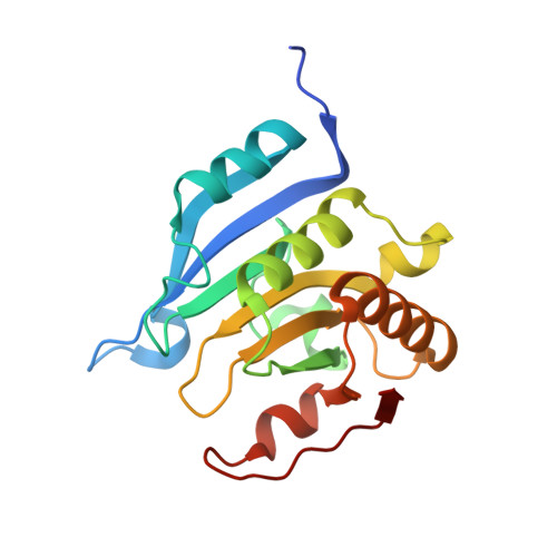
Structural insights into parasite EIF4E binding specificity for m7G and m2,2,7G mRNA cap.

Liu, W.Zhao, R.McFarland, C.Kieft, J.Niedzwiecka, A.Jankowska-Anyszka, M.Stepinski, J.Darzynkiewicz, E.Jones, D.N.Davis, R.E.

(2009) J Biol Chem 284: 31336-31349

  • DOI: https://doi.org/10.1074/jbc.M109.049858
  • Primary Citation of Related Structures:  
    3HXG, 3HXI

  • PubMed Abstract: 

    The eukaryotic translation initiation factor eIF4E recognizes the mRNA cap, a key step in translation initiation. Here we have characterized eIF4E from the human parasite Schistosoma mansoni. Schistosome mRNAs have either the typical monomethylguanosine (m(7)G) or a trimethylguanosine (m(2,2,7)G) cap derived from spliced leader trans-splicing. Quantitative fluorescence titration analyses demonstrated that schistosome eIF4E has similar binding specificity for both caps. We present the first crystal structure of an eIF4E with similar binding specificity for m(7)G and m(2,2,7)G caps. The eIF4E.m(7)GpppG structure demonstrates that the schistosome protein binds monomethyl cap in a manner similar to that of single specificity eIF4Es and exhibits a structure similar to other known eIF4Es. The structure suggests an alternate orientation of a conserved, key Glu-90 in the cap-binding pocket that may contribute to dual binding specificity and a position for mRNA bound to eIF4E consistent with biochemical data. Comparison of NMR chemical shift perturbations in schistosome eIF4E on binding m(7)GpppG and m(2,2,7)GpppG identified key differences between the two complexes. Isothermal titration calorimetry demonstrated significant thermodynamics differences for the binding process with the two caps (m(7)G versus m(2,2,7)G). Overall the NMR and isothermal titration calorimetry data suggest the importance of intrinsic conformational flexibility in the schistosome eIF4E that enables binding to m(2,2,7)G cap.


  • Organizational Affiliation

    Department of Biochemistry and Molecular Genetics, University of Colorado School of Medicine, Aurora, Colorado 80045, USA.


Macromolecules
Find similar proteins by:  (by identity cutoff)  |  3D Structure
Entity ID: 1
MoleculeChains Sequence LengthOrganismDetailsImage
Eukaryotic Translation Initiation Factor 4E189Schistosoma mansoniMutation(s): 0 
Gene Names: eif4e
Entity Groups  
Sequence Clusters30% Identity50% Identity70% Identity90% Identity95% Identity100% Identity
Sequence Annotations
Expand
  • Reference Sequence

Find similar proteins by:  Sequence   |   3D Structure  

Entity ID: 2
MoleculeChains Sequence LengthOrganismDetailsImage
Eukaryotic translation initiation factor 4E-binding protein 1B [auth C]21Homo sapiensMutation(s): 0 
UniProt & NIH Common Fund Data Resources
Find proteins for Q13541 (Homo sapiens)
Explore Q13541 
Go to UniProtKB:  Q13541
PHAROS:  Q13541
GTEx:  ENSG00000187840 
Entity Groups  
Sequence Clusters30% Identity50% Identity70% Identity90% Identity95% Identity100% Identity
UniProt GroupQ13541
Sequence Annotations
Expand
  • Reference Sequence
Small Molecules
Ligands 1 Unique
IDChains Name / Formula / InChI Key2D Diagram3D Interactions
GTA
Query on GTA

Download Ideal Coordinates CCD File 
C [auth A]P1-7-METHYLGUANOSINE-P3-ADENOSINE-5',5'-TRIPHOSPHATE
C21 H30 N10 O17 P3
QQOHNVHGNZYSBP-XPWFQUROSA-O
Binding Affinity Annotations 
IDSourceBinding Affinity
GTA Binding MOAD:  3HXG Kd: 270 (nM) from 1 assay(s)
Experimental Data & Validation

Experimental Data

  • Method: X-RAY DIFFRACTION
  • Resolution: 2.10 Å
  • R-Value Free: 0.269 
  • R-Value Work: 0.210 
  • R-Value Observed: 0.215 
  • Space Group: P 21 21 2
Unit Cell:
Length ( Å )Angle ( ˚ )
a = 45.56α = 90
b = 125.4β = 90
c = 37.34γ = 90
Software Package:
Software NamePurpose
PHASESphasing
REFMACrefinement
d*TREKdata reduction
d*TREKdata scaling

Structure Validation

View Full Validation Report


Ligand Structure Quality Assessment 


Entry History 

Deposition Data

Revision History  (Full details and data files)

  • Version 1.0: 2009-08-25
    Type: Initial release
  • Version 1.1: 2011-07-13
    Changes: Version format compliance
  • Version 1.2: 2023-09-06
    Changes: Data collection, Database references, Derived calculations, Refinement description