3GTT

Mouse SOD1


Experimental Data Snapshot

  • Method: X-RAY DIFFRACTION
  • Resolution: 2.40 Å
  • R-Value Free: 0.197 
  • R-Value Work: 0.158 
  • R-Value Observed: 0.160 

wwPDB Validation   3D Report Full Report


This is version 1.2 of the entry. See complete history


Literature

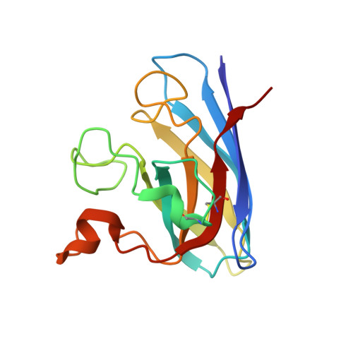
Structures of mouse SOD1 and human/mouse SOD1 chimeras.

Seetharaman, S.V.Taylor, A.B.Holloway, S.Hart, P.J.

(2010) Arch Biochem Biophys 503: 183-190

  • DOI: https://doi.org/10.1016/j.abb.2010.08.014
  • Primary Citation of Related Structures:  
    3GTT, 3GTV, 3LTV

  • PubMed Abstract: 

    Mutations in human copper-zinc superoxide dismutase (SOD1) cause an inherited form of amyotrophic lateral sclerosis (ALS). Inclusions enriched in pathogenic SOD1 accumulate in the spinal cords of transgenic mice expressing these proteins, but endogenous mouse SOD1 is not found as a component of these aggregates. In the accompanying paper, Karch and colleagues analyze aggregation propensities of human/mouse SOD1 chimeras in cell culture and identify two sequence elements in the human enzyme that seem to enhance its aggregation relative to the mouse enzyme. Here, we report the first structure of mouse SOD1 along with those of SOD1 chimeras in which residues 1-80 come from human SOD1 and residues 81-153 come from mouse SOD1 and vice versa. Taken together, the structural and cell-based data suggest a model in which residues Q42 and Q123 in mouse SOD1 modulate non-native SOD1-SOD1 intermolecular interactions at edge strands in the SOD1 Greek key β-barrel.


  • Organizational Affiliation

    Department of Biochemistry, The University of Texas Health Science Center, San Antonio, 78229, USA.


Macromolecules
Find similar proteins by:  (by identity cutoff)  |  3D Structure
Entity ID: 1
MoleculeChains Sequence LengthOrganismDetailsImage
Superoxide dismutase [Cu-Zn]
A, B, C, D, E
A, B, C, D, E, F
153Mus musculusMutation(s): 0 
Gene Names: Sod1
EC: 1.15.1.1
UniProt
Find proteins for P08228 (Mus musculus)
Explore P08228 
Go to UniProtKB:  P08228
Entity Groups  
Sequence Clusters30% Identity50% Identity70% Identity90% Identity95% Identity100% Identity
UniProt GroupP08228
Sequence Annotations
Expand
  • Reference Sequence
Experimental Data & Validation

Experimental Data

  • Method: X-RAY DIFFRACTION
  • Resolution: 2.40 Å
  • R-Value Free: 0.197 
  • R-Value Work: 0.158 
  • R-Value Observed: 0.160 
  • Space Group: C 2 2 21
Unit Cell:
Length ( Å )Angle ( ˚ )
a = 112.454α = 90
b = 194.495β = 90
c = 149.841γ = 90
Software Package:
Software NamePurpose
CrystalCleardata collection
PHASERphasing
PHENIXrefinement
HKL-2000data reduction
HKL-2000data scaling

Structure Validation

View Full Validation Report


Entry History 

Deposition Data

Revision History  (Full details and data files)

  • Version 1.0: 2010-09-08
    Type: Initial release
  • Version 1.1: 2011-07-13
    Changes: Version format compliance
  • Version 1.2: 2023-09-06
    Changes: Data collection, Database references, Derived calculations, Refinement description