3GNI

Structure of STRAD and MO25


Experimental Data Snapshot

  • Method: X-RAY DIFFRACTION
  • Resolution: 2.35 Å
  • R-Value Free: 0.254 
  • R-Value Work: 0.206 
  • R-Value Observed: 0.207 

wwPDB Validation   3D Report Full Report


Ligand Structure Quality Assessment 


This is version 1.2 of the entry. See complete history


Literature

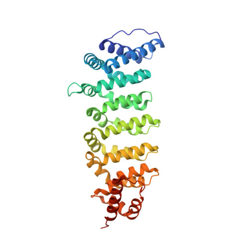
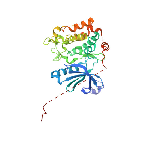
ATP and MO25alpha regulate the conformational state of the STRADalpha pseudokinase and activation of the LKB1 tumour suppressor

Zeqiraj, E.Filippi, B.M.Goldie, S.Navratilova, I.Boudeau, J.Deak, M.Alessi, D.R.van Aalten, D.M.

(2009) PLoS Biol 7: e1000126-e1000126

  • DOI: https://doi.org/10.1371/journal.pbio.1000126
  • Primary Citation of Related Structures:  
    3GNI

  • PubMed Abstract: 

    Pseudokinases lack essential residues for kinase activity, yet are emerging as important regulators of signal transduction networks. The pseudokinase STRAD activates the LKB1 tumour suppressor by forming a heterotrimeric complex with LKB1 and the scaffolding protein MO25. Here, we describe the structure of STRADalpha in complex with MO25alpha. The structure reveals an intricate web of interactions between STRADalpha and MO25alpha involving the alphaC-helix of STRADalpha, reminiscent of the mechanism by which CDK2 interacts with cyclin A. Surprisingly, STRADalpha binds ATP and displays a closed conformation and an ordered activation loop, typical of active protein kinases. Inactivity is accounted for by nonconservative substitution of almost all essential catalytic residues. We demonstrate that binding of ATP enhances the affinity of STRADalpha for MO25alpha, and conversely, binding of MO25alpha promotes interaction of STRADalpha with ATP. Mutagenesis studies reveal that association of STRADalpha with either ATP or MO25alpha is essential for LKB1 activation. We conclude that ATP and MO25alpha cooperate to maintain STRADalpha in an "active" closed conformation required for LKB1 activation. It has recently been demonstrated that a mutation in human STRADalpha that truncates a C-terminal region of the pseudokinase domain leads to the polyhydramnios, megalencephaly, symptomatic epilepsy (PMSE) syndrome. We demonstrate this mutation destabilizes STRADalpha and prevents association with LKB1. In summary, our findings describe one of the first structures of a genuinely inactive pseudokinase. The ability of STRADalpha to activate LKB1 is dependent on a closed "active" conformation, aided by ATP and MO25alpha binding. Thus, the function of STRADalpha is mediated through an active kinase conformation rather than kinase activity. It is possible that other pseudokinases exert their function through nucleotide binding and active conformations.


  • Organizational Affiliation

    Division of Molecular Microbiology, College of Life Sciences, University of Dundee, Dundee, Scotland.


Macromolecules
Find similar proteins by:  (by identity cutoff)  |  3D Structure
Entity ID: 1
MoleculeChains Sequence LengthOrganismDetailsImage
Protein Mo25341Homo sapiensMutation(s): 0 
Gene Names: CAB39MO25CGI-66
UniProt & NIH Common Fund Data Resources
Find proteins for Q9Y376 (Homo sapiens)
Explore Q9Y376 
Go to UniProtKB:  Q9Y376
PHAROS:  Q9Y376
GTEx:  ENSG00000135932 
Entity Groups  
Sequence Clusters30% Identity50% Identity70% Identity90% Identity95% Identity100% Identity
UniProt GroupQ9Y376
Sequence Annotations
Expand
  • Reference Sequence
Find similar proteins by:  (by identity cutoff)  |  3D Structure
Entity ID: 2
MoleculeChains Sequence LengthOrganismDetailsImage
STRAD alpha389Homo sapiensMutation(s): 0 
Gene Names: STRADALYK5STRAD
UniProt & NIH Common Fund Data Resources
Find proteins for Q7RTN6 (Homo sapiens)
Explore Q7RTN6 
Go to UniProtKB:  Q7RTN6
PHAROS:  Q7RTN6
GTEx:  ENSG00000266173 
Entity Groups  
Sequence Clusters30% Identity50% Identity70% Identity90% Identity95% Identity100% Identity
UniProt GroupQ7RTN6
Sequence Annotations
Expand
  • Reference Sequence
Small Molecules
Ligands 2 Unique
IDChains Name / Formula / InChI Key2D Diagram3D Interactions
ATP
Query on ATP

Download Ideal Coordinates CCD File 
D [auth B]ADENOSINE-5'-TRIPHOSPHATE
C10 H16 N5 O13 P3
ZKHQWZAMYRWXGA-KQYNXXCUSA-N
CIT
Query on CIT

Download Ideal Coordinates CCD File 
C [auth A]CITRIC ACID
C6 H8 O7
KRKNYBCHXYNGOX-UHFFFAOYSA-N
Binding Affinity Annotations 
IDSourceBinding Affinity
ATP Binding MOAD:  3GNI Kd: 20 (nM) from 1 assay(s)
Experimental Data & Validation

Experimental Data

  • Method: X-RAY DIFFRACTION
  • Resolution: 2.35 Å
  • R-Value Free: 0.254 
  • R-Value Work: 0.206 
  • R-Value Observed: 0.207 
  • Space Group: P 21 21 21
Unit Cell:
Length ( Å )Angle ( ˚ )
a = 73.678α = 90
b = 82.874β = 90
c = 134.292γ = 90
Software Package:
Software NamePurpose
DNAdata collection
MOLREPphasing
REFMACrefinement
MOSFLMdata reduction
SCALAdata scaling

Structure Validation

View Full Validation Report


Ligand Structure Quality Assessment 


Entry History 

Deposition Data

Revision History  (Full details and data files)

  • Version 1.0: 2009-06-16
    Type: Initial release
  • Version 1.1: 2011-07-13
    Changes: Advisory, Refinement description, Version format compliance
  • Version 1.2: 2023-09-06
    Changes: Data collection, Database references, Derived calculations, Refinement description