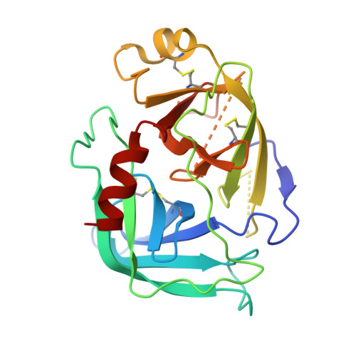
Structure of granzyme C reveals an unusual mechanism of protease autoinhibition
Kaiserman, D., Buckle, A.M., Van Damme, P., Irving, J.A., Law, R.H.P., Matthews, A.Y., Bashtannyk-Puhalovich, T., Langendorf, C., Thompson, P., Vandekerckhove, J., Gevaert, K., Whisstock, J.C., Bird, P.I.(2009) Proc Natl Acad Sci U S A 106: 5587-5592
- PubMed: 19299505
- DOI: https://doi.org/10.1073/pnas.0811968106
- Primary Citation of Related Structures:
3FZZ, 3G01 - PubMed Abstract:
Proteases act in important homeostatic pathways and are tightly regulated. Here, we report an unusual structural mechanism of regulation observed by the 2.5-A X-ray crystal structure of the serine protease, granzyme C. Although the active-site triad residues adopt canonical conformations, the oxyanion hole is improperly formed, and access to the primary specificity (S1) pocket is blocked through a reversible rearrangement involving Phe-191. Specifically, a register shift in the 190-strand preceding the active-site serine leads to Phe-191 filling the S1 pocket. Mutation of a unique Glu-Glu motif at positions 192-193 unlocks the enzyme, which displays chymase activity, and proteomic analysis confirms that activity of the wild-type protease can be released through interactions with an appropriate substrate. The 2.5-A structure of the unlocked enzyme reveals unprecedented flexibility in the 190-strand preceding the active-site serine that results in Phe-191 vacating the S1 pocket. Overall, these observations describe a broadly applicable mechanism of protease regulation that cannot be predicted by template-based modeling or bioinformatic approaches alone.
Organizational Affiliation:
Department of Biochemistry and Molecular Biology, Monash University, Clayton 3800, Australia.