3FUG

Crystal Structure of the Retinoid X Receptor Ligand Binding Domain Bound to the Synthetic Agonist 3-[4-Hydroxy-3-(3,5,5,8,8-pentamethyl-5,6,7,8-tetrahydronaphthalen-2-yl)-phenyl]acrylic Acid


Experimental Data Snapshot

  • Method: X-RAY DIFFRACTION
  • Resolution: 2.00 Å
  • R-Value Free: 0.241 
  • R-Value Work: 0.205 
  • R-Value Observed: 0.207 

wwPDB Validation   3D Report Full Report


Ligand Structure Quality Assessment 


This is version 1.2 of the entry. See complete history


Literature

Modulating retinoid X receptor with a series of (E)-3-[4-hydroxy-3-(3-alkoxy-5,5,8,8-tetramethyl-5,6,7,8-tetrahydronaphthalen-2-yl)phenyl]acrylic acids and their 4-alkoxy isomers.

Perez Santin, E.Germain, P.Quillard, F.Khanwalkar, H.Rodriguez-Barrios, F.Gronemeyer, H.de Lera, A.R.Bourguet, W.

(2009) J Med Chem 52: 3150-3158

  • DOI: https://doi.org/10.1021/jm900096q
  • Primary Citation of Related Structures:  
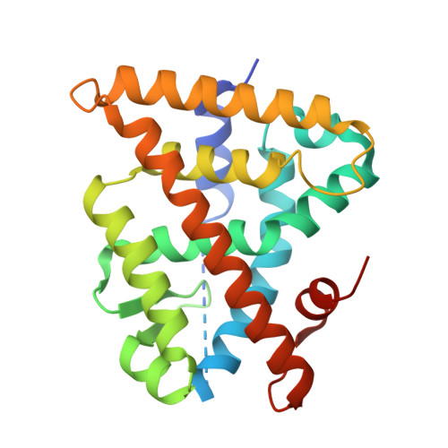
    3FUG

  • PubMed Abstract: 

    Rexinoids are ligands for the retinoid X receptor (RXR) that have great promise for both the prevention and treatment of cancer and metabolic diseases. In this regard, synthetic, functional, and structural investigations into the structure-activity relationships of derivatives of the potent RXR agonist (E)-3-[3-(3,5,5,8,8-pentamethyl-5,6,7,8-tetrahydronaphthalen-2-yl)-4-hydroxyphenyl]acrylic acid (CD3254, 9) have been conducted. We recently reported on the characterization of a series of C3'-substituted alkyl ether analogues of 9 (10a-f), which display activities ranging from partial agonists to pure antagonists. The importance of the position of the alkoxy side chain for ligand activity has been further explored with the synthesis of C4'-substituted analogues (11a-f). Here we describe the synthesis of compounds 11a-f, which appear functionally different from their isomeric counterparts, as judged from transactivation assays and fluorescence anisotropy experiments. We also report on the 2.0 A resolution structure of RXR in complex with the parent compound 9, which helps understanding of the impact of the alkyl side chain location on ligand activity.


  • Organizational Affiliation

    Departamento de Química Orgánica, Facultad de Química, Universidade de Vigo, 36310 Vigo, Spain.


Macromolecules
Find similar proteins by:  (by identity cutoff)  |  3D Structure
Entity ID: 1
MoleculeChains Sequence LengthOrganismDetailsImage
Retinoic acid receptor RXR-alpha240Homo sapiensMutation(s): 0 
Gene Names: NR2B1RXRA
UniProt & NIH Common Fund Data Resources
Find proteins for P19793 (Homo sapiens)
Explore P19793 
Go to UniProtKB:  P19793
PHAROS:  P19793
GTEx:  ENSG00000186350 
Entity Groups  
Sequence Clusters30% Identity50% Identity70% Identity90% Identity95% Identity100% Identity
UniProt GroupP19793
Sequence Annotations
Expand
  • Reference Sequence

Find similar proteins by:  Sequence   |   3D Structure  

Entity ID: 2
MoleculeChains Sequence LengthOrganismDetailsImage
Nuclear receptor coactivator 213N/AMutation(s): 0 
UniProt & NIH Common Fund Data Resources
Find proteins for Q15596 (Homo sapiens)
Explore Q15596 
Go to UniProtKB:  Q15596
PHAROS:  Q15596
GTEx:  ENSG00000140396 
Entity Groups  
Sequence Clusters30% Identity50% Identity70% Identity90% Identity95% Identity100% Identity
UniProt GroupQ15596
Sequence Annotations
Expand
  • Reference Sequence
Small Molecules
Ligands 1 Unique
IDChains Name / Formula / InChI Key2D Diagram3D Interactions
2E3
Query on 2E3

Download Ideal Coordinates CCD File 
C [auth A](2E)-3-[4-hydroxy-3-(3,5,5,8,8-pentamethyl-5,6,7,8-tetrahydronaphthalen-2-yl)phenyl]prop-2-enoic acid
C24 H28 O3
DYLLZSVPAUUSSB-VQHVLOKHSA-N
Experimental Data & Validation

Experimental Data

  • Method: X-RAY DIFFRACTION
  • Resolution: 2.00 Å
  • R-Value Free: 0.241 
  • R-Value Work: 0.205 
  • R-Value Observed: 0.207 
  • Space Group: P 43 21 2
Unit Cell:
Length ( Å )Angle ( ˚ )
a = 65.763α = 90
b = 65.763β = 90
c = 110.857γ = 90
Software Package:
Software NamePurpose
MOSFLMdata reduction
SCALAdata scaling
MOLREPphasing
REFMACrefinement
PDB_EXTRACTdata extraction
DNAdata collection

Structure Validation

View Full Validation Report


Ligand Structure Quality Assessment 


Entry History 

Deposition Data

  • Released Date: 2009-05-12 
  • Deposition Author(s): Bourguet, W.

Revision History  (Full details and data files)

  • Version 1.0: 2009-05-12
    Type: Initial release
  • Version 1.1: 2011-07-13
    Changes: Version format compliance
  • Version 1.2: 2023-09-06
    Changes: Data collection, Database references, Derived calculations, Refinement description