3FJO

Structure of chimeric YH CPR


Experimental Data Snapshot

  • Method: X-RAY DIFFRACTION
  • Resolution: 2.50 Å
  • R-Value Free: 0.290 
  • R-Value Work: 0.222 
  • R-Value Observed: 0.225 

wwPDB Validation   3D Report Full Report


Ligand Structure Quality Assessment 


This is version 1.3 of the entry. See complete history


Literature

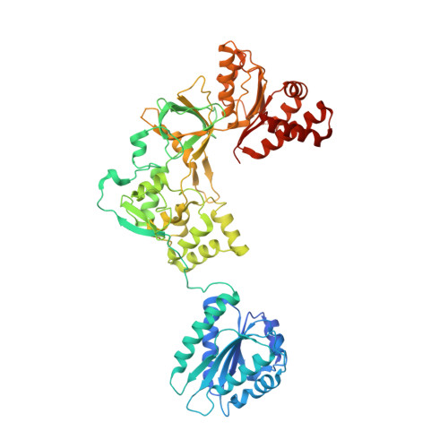
Structure of the open conformation of a functional chimeric NADPH cytochrome P450 reductase

Aigrain, L.Pompon, D.Morera, S.Truan, G.

(2009) EMBO Rep 10: 742-747

  • DOI: https://doi.org/10.1038/embor.2009.82
  • Primary Citation of Related Structures:  
    3FJO

  • PubMed Abstract: 

    Two catalytic domains, bearing FMN and FAD cofactors, joined by a connecting domain, compose the core of the NADPH cytochrome P450 reductase (CPR). The FMN domain of CPR mediates electron shuttling from the FAD domain to cytochromes P450. Together, both enzymes form the main mixed-function oxidase system that participates in the metabolism of endo- and xenobiotic compounds in mammals. Available CPR structures show a closed conformation, with the two cofactors in tight proximity, which is consistent with FAD-to-FMN, but not FMN-to-P450, electron transfer. Here, we report the 2.5 A resolution crystal structure of a functionally competent yeast-human chimeric CPR in an open conformation, compatible with FMN-to-P450 electron transfer. Comparison with closed structures shows a major conformational change separating the FMN and FAD cofactors from 86 A.


  • Organizational Affiliation

    Centre de Génétique Moléculaire, FRE3144, Gif-sur-Yvette, France.


Macromolecules
Find similar proteins by:  (by identity cutoff)  |  3D Structure
Entity ID: 1
MoleculeChains Sequence LengthOrganismDetailsImage
NADPH-cytochrome P450 reductase637Saccharomyces cerevisiaeHomo sapiens
This entity is chimeric
Mutation(s): 0 
Gene Names: NCP1POR
EC: 1.6.2.4
UniProt & NIH Common Fund Data Resources
Find proteins for P16603 (Saccharomyces cerevisiae (strain ATCC 204508 / S288c))
Explore P16603 
Go to UniProtKB:  P16603
Find proteins for P16435 (Homo sapiens)
Explore P16435 
Go to UniProtKB:  P16435
PHAROS:  P16435
GTEx:  ENSG00000127948 
Entity Groups  
Sequence Clusters30% Identity50% Identity70% Identity90% Identity95% Identity100% Identity
UniProt GroupsP16435P16603
Sequence Annotations
Expand
  • Reference Sequence
Small Molecules
Ligands 2 Unique
IDChains Name / Formula / InChI Key2D Diagram3D Interactions
FAD
Query on FAD

Download Ideal Coordinates CCD File 
C [auth A]FLAVIN-ADENINE DINUCLEOTIDE
C27 H33 N9 O15 P2
VWWQXMAJTJZDQX-UYBVJOGSSA-N
FMN
Query on FMN

Download Ideal Coordinates CCD File 
B [auth A]FLAVIN MONONUCLEOTIDE
C17 H21 N4 O9 P
FVTCRASFADXXNN-SCRDCRAPSA-N
Experimental Data & Validation

Experimental Data

  • Method: X-RAY DIFFRACTION
  • Resolution: 2.50 Å
  • R-Value Free: 0.290 
  • R-Value Work: 0.222 
  • R-Value Observed: 0.225 
  • Space Group: C 1 2 1
Unit Cell:
Length ( Å )Angle ( ˚ )
a = 146.8α = 90
b = 60.7β = 103.36
c = 78.86γ = 90
Software Package:
Software NamePurpose
ADSCdata collection
PHASERphasing
REFMACrefinement
XDSdata reduction
XDSdata scaling

Structure Validation

View Full Validation Report


Ligand Structure Quality Assessment 


Entry History 

Deposition Data

Revision History  (Full details and data files)

  • Version 1.0: 2009-06-16
    Type: Initial release
  • Version 1.1: 2011-07-13
    Changes: Advisory, Version format compliance
  • Version 1.2: 2017-08-16
    Changes: Refinement description, Source and taxonomy
  • Version 1.3: 2023-11-01
    Changes: Data collection, Database references, Derived calculations, Refinement description